Surface Modifications of Titanium Plates to Enhance Osteogenesis: Advances, Challenges, and Clinical Insights
-
Randy Bachelard Nziengui Raby
Institute of Biomedical Engineering, Central South University, Yuelu District, Changsha 410011, Hunan, People’s Republic of China
Guy Armel Bounda

Section Académique, Association des Etudiants et Stagiaires Gabonais de Chine, Beijing 100600, People’s Republic of China
Kelvin Stefel Lebila MoutsiDepartment of Radiology, The Second Xiangya Hospital, Central South University, 139# Middle Renmin Road, Changsha 410011, Hunan, People’s Republic of China
Idriss François Ntsame AllogoSchool of Public Health, Central South University, Yuelu District, Changsha 410011, Hunan, People’s Republic of China
Justia Varaine Mengue ObameSection Académique, Association des Etudiants et Stagiaires Gabonais de Chine, Beijing 100600, People’s Republic of China
Monique Lauriane Adou OvoloAnhui Polytechnic University, Beijing Middle Road, Wuhu 241000, Anhui, People’s Republic of China
Kwensy Djoyce OndoDepartment of Orthopaedics and Traumatology, Longhua Hospital Affiliated to Shanghai University of Traditional Chinese Medicine, Pudong District Shangnan Road 1000 Longshang Gangercun 45#, Shanghai, People’s Republic of China
Geraldine Esther AwakossaCancer Center, Renmin Hospital of Wuhan University, Wuhan, 430060, People’s Republic of China
Kiny-Binyumbe DoukagaDepartment of Gynecology and Obstetrics, Shandong First Medical University, 619# Great Wall Road Beijipo Street, Daiyue District, Tai'an, Shandong, People’s Republic of China
Rosanna Tryphene Massounga MayomboInstitute of Hematology, Union Hospital, Tongji Medical College, Huazhong University of Science and Technology, 1277# Jiefang Avenue, Wuhan 430022, Hubei, People’s Republic of China
Dally Mengue Bea MedzangDepartment of Internal Medicine, The Third Xiangya Hospital, Central South University, 139# Middle Renmin Road, Changsha 410011, Hunan, People’s Republic of China
Marie Mical Tsamba MpiraDepartment of Internal Medicine, The Third Xiangya Hospital, Central South University, 139# Middle Renmin Road, Changsha 410011, Hunan, People’s Republic of China
Maywann BengonoZhejiang University of Traditional Chinese Medicine, 548# Binwen Road, Binjiang District, Hangzhou, Zhejiang, People’s Republic of China
Ulrich Freddy Nziengui MbadingaDepartment of Food Science and Engineering, Changsha University of Science and Technology, 960, 2" Section, Wanjiali South RD, Changsha 410004, Hunan, People’s Republic of China
Maëlle Noémie Mengue ObameDepartment of Pharmacy, Linyi University, Middle of Shuangling Road, Linyi City 410004, Shandong, People’s Republic of China
Noreen Teingue DodoDepartment of Gynecology and Obstetrics, Dalian Medical University, 9 West Section, Lvshun South Road, Dalian 116041, Liaoning, People’s Republic of China
Zhi HuangInstitute of Biomedical Engineering, Central South University, Yuelu District, Changsha 410011, Hunan, People’s Republic of China
| Received 21 Apr, 2025 |
Accepted 20 Jun, 2025 |
Published 21 Jun, 2025 |
Titanium and its alloys are indispensable in bone regeneration and implantology due to their superior biocompatibility and mechanical strength. This review synthesizes recent advances in titanium plate surface modifications for osteogenesis, focusing on innovative approaches in surface engineering, such as the integration of nanomaterials, bioactive coatings, and laser treatments, to enhance osseointegration, antibacterial properties, and implant durability. A systematic literature search (2010-2024) was conducted across PubMed, Web of Science, Scopus, and ScienceDirect to comprehensively capture high-quality studies in this field. A critical assessment places these modifications within the context of improved clinical outcomes in orthopedic and reconstructive surgeries, addressing challenges of long-term stability, cost-effectiveness, and regulatory compliance. Overall, this comprehensive analysis provides valuable insights into current practices and outlines future research directions essential for optimizing implant performance, aligning with the scope of challenges by presenting innovative solutions in the rapidly evolving field of biomaterials.
| Copyright © 2025 Raby et al. This is an open-access article distributed under the Creative Commons Attribution License, which permits unrestricted use, distribution, and reproduction in any medium, provided the original work is properly cited. |
INTRODUCTION
The quest for ideal materials in bone regeneration and implantation has led to the widespread adoption of titanium and its alloys. Their biocompatibility, high strength-to-weight ratio, corrosion resistance, and nonmagnetic properties make them excellent candidates for medical implants1-3. The phenomenon of osseointegration, first described by Professor Brånemark, wherein a stable combination of titanium and bone tissue is achieved, has further cemented titanium’s position in dentistry and orthopedics3-5. However, while titanium offers remarkable advantages, its bio-inert nature necessitates surface modifications to enhance its interaction with biological tissues and to promote optimal bone regeneration3.
Titanium implants success is tied to their surface characteristics, which influence cell adhesion, proliferation, and differentiation6. These surface properties are not just about topography, but also chemistry and wettability7. In recent years, there has been an increasing focus on modifying the surface of titanium implants to enhance their biological properties and improve clinical outcomes3. This has led to the development of a range of surface modification techniques aimed at enhancing osteointegration and promoting a healthier tissue environment7. This review aims to provide a structured insight into the current state of titanium plate surface technology in osteogenesis, highlighting the advancements, identifying challenges, projecting future possibilities, and providing essential recommendations for research and clinical practice.
Methodology: A comprehensive and systematic literature search was conducted utilizing several electronic databases, including PubMed, Web of Science, Scopus, and ScienceDirect, covering publications from 2010 to 2024. This search encompassed original research articles, review articles, and case studies pertinent to the clinical applications of titanium plate surfaces in osteogenesis. To further expand the search, Google Scholar was also queried to identify studies not indexed in the primary databases. A combination of keywords such as “titanium implants”, “osteogenesis”, “surface modification”, “osseointegration”, “antibacterial coatings”, “bone regeneration”, “3D-printed titanium”, “laser treatment titanium”, and “clinical applications” was employed to ensure comprehensive coverage of the topic. Only studies published in English were included in this review. The selection of articles was based on their relevance to the clinical applications of titanium plate surfaces, with a particular emphasis on scientific rigor and the practical implications for osteogenesis.
Breakthroughs of clinical application of titanium plate surface in osteogenesis: Over the past decades, the clinical use of titanium in osteogenesis has seen significant breakthroughs driven by advancements in material science, surface engineering, and a deeper understanding of biological interactions.
Surface modification and enhanced osteointegration: Surface modification of titanium implants is critical for improving osseointegration, directly influencing the success of orthopedic and dental implants2,7. Techniques such as sandblasting, acid etching (SLA), micro-arc oxidation (MAO), laser treatments, and nanotopography have been developed to enhance cell adhesion, proliferation, and differentiation on titanium surfaces3,7,8. They offer significant improvements in both initial biological response and long-term implant stability.
The SLA, a widely used method, creates a micro-rough surface that increases surface area, promoting osteoblast adhesion and bone formation9. MAO generates a porous oxide layer, which can be enriched with bioactive elements, fostering a favorable environment for bone healing3. Laser treatments offer precise micro and nanoscale surface modifications that improve cell alignment and osteogenic differentiation, without contaminating the surface8,10.
Nanotopographical modifications, such as TiO2 nanotubes, enhance osseointegration by improving protein adsorption and facilitating osteoblast function11. These nanoscale features also promote stem cell differentiation, making them a promising strategy for better implant integration2,7,12.
Enhanced biocompatibility through surface chemistry: Optimizing titanium implants through both surface chemistry and topographical modifications is emerging as a critical strategy to enhance osseointegration and expand the applications of these devices in skeletal reconstruction and regenerative medicine. The incorporation of bioactive ions, multifunctional coatings, and innovative treatments, such as Recombinant Human Bone Morphogenetic Protein-2 (rhBMP-2) and Semaphorin 3A (Sema3A), has shown considerable promise in improving clinical outcomes.
Surface chemistry modifications are essential for enhancing biocompatibility and promoting bone integration. Recent advances have emphasized the use of bioactive ions like calcium, phosphate, and magnesium, which facilitate hydroxyapatite formation on the implant surface a process fundamental to robust bone integration3,7. Calcium, in particular, plays a pivotal role by stimulating bone attachment and growth, thereby improving endosseous integration13.
In addition to chemical modifications, the surface topography of titanium implants significantly influences osseointegration. Techniques such as erbium-doped yttrium aluminum garnet (Er:YAG) laser irradiation have been shown to enhance osteoblast proliferation and cell attachment, underscoring the importance of surface treatments in achieving favorable clinical outcomes14. Furthermore, combining topographical modifications with chemical approaches for example, through chitosan/hydroxyapatite coatings can further boost osteoblast differentiation and overall implant stability15. Dual-modified surfaces that integrate both chemical and topographical features have been shown to promote cellular behavior and facilitate tissue regeneration15, with factors like Sema3A enhancing osteogenesis, particularly in osteoporotic models17-19.
Pharmacological interventions also contribute to the optimization of implant performance. For instance, while Etoricoxib a Cyclooxygenase-2 (COX-2) inhibitor effectively serves as an analgesic and mitigates osteoclastogenesis, periodontal bone loss, and root resorption during orthodontic tooth movement, it does not affect the movement itself20. In contrast, sclerostin antibody (Scl-Ab) has demonstrated efficacy in enhancing implant fixation and bone formation in osteoporotic conditions21. Moreover, nanotubular structures loaded with strontium (Sr) and silver (Ag) have improved both the biological and mechanical properties of titanium surfaces by providing antibacterial benefits and supporting bone repair22. Innovative strategies, including the functionalization of titanium with amorphous calcium phosphate and the immobilization of rhBMP-2, have further advanced the field by enhancing osteogenic differentiation and bone formation23,24. In challenging clinical scenarios, even brief applications of mechanical stress, such as two weeks of applied force, can stimulate bone formation, potentially offering new solutions for accelerating tooth movement in the maxillary sinus25. Additionally, titanium implants with nano-pillared topographies and laser-sintered Ti-6Al-4V designs have been shown to support enhanced tissue integration and bone growth26.
Recent developments in immobilization techniques, such as anchoring rhBMP-2 via nano-anchored oligonucleotide strands, have demonstrated promising results in maintaining the bioactivity of the growth factor under standard sterilization conditions, thereby ensuring sustained osteogenic stimulation and improved bone-implant contact5,27. The naturally occurring Titanium Dioxide (TiO2) layer also plays a crucial role by providing chemical stability and promoting biocompatibility2, while hydrophilic surface modifications further enhance protein adsorption critical for osteoblast adhesion and proliferation7,28.
Addressing the significant clinical challenge posed by bacterial infections28,29, particularly by drug-resistant strains like Methicillin-resistant Staphylococcus aureus (MRSA), which readily form antibiotic-resistant biofilms on biomaterials5,29. Advanced surface modifications are incorporating sophisticated controlled-release strategies for potent antibiotics such as vancomycin29. These strategies leverage stimulus-responsive mechanisms, allowing for targeted drug release triggered by specific cues present at the infection site28. Examples include platforms utilizing magnetic nanoparticles like Fe3O4@chitosan5,28, which can be engineered for magnetic guidance and subsequent drug release stimulated by an alternating magnetic field (AMF). Another approach involves employing linkers, such as 4,4'-azobis (4-cyanovaleric acid) (ACVA)29, capable of facilitating drug release in response to reactive oxygen species (ROS)-molecules abundant in infected wound microenvironments. By integrating these stimuli-responsive controlled-release systems onto titanium implant surfaces, it becomes possible to deliver therapeutic agents like vancomycin locally and on-demand, potentially enhancing both infection eradication and the overall osseointegration process28,29.
While traditional methods have limitations30-32, promising new strategies are emerging to directly prevent bacterial adhesion and growth on implants. For instance, research into materials like polyvinyl alcohol (PVA) for coating titanium implants shows potential to effectively inhibit bacterial growth and biofilm formation, as demonstrated in both laboratory and animal studies33. However, ensuring sustained antibacterial efficacy and long-term biocompatibility of such novel coatings in the complex biological environment remains a key area for research and validation. Finally, the application of biomimetic materials and advanced fabrication techniques offers a promising avenue for bone tissue regeneration24. By replicating the natural bone microenvironment, these approaches foster more efficient healing and accelerate regenerative processes, ultimately improving overall implant integration34-36.
Collectively, these multifaceted advancements illustrate the promising future of titanium implant modifications, offering new avenues for enhanced clinical outcomes in both skeletal reconstruction and regenerative medicine.
Antibacterial innovations: Implant-related infections remain a major challenge in orthopedic surgery, complicating the success of titanium implants and prolonging recovery. Recent advancements in antibacterial coatings aim to prevent bacterial adhesion, biofilm formation, and infections, thereby enhancing implant stability. One notable development is iodine-doped Titanium Dioxide (TiO2) nanotubes, which exhibit enhanced antimicrobial properties, particularly against Staphylococcus aureus, while promoting biocompatibility and osseointegration37.
Silver nanoparticle coatings have also gained attention for their potent antibacterial effects, preventing biofilm formation. However, regulatory concerns regarding the use of fluorine-based polymers in these coatings require the development of safer alternatives1,38.
These regulatory hurdles necessitate the development of safer, more biocompatible alternatives that maintain the antimicrobial efficacy of silver while addressing safety concerns. Innovative approaches, such as slippery liquid-infused porous surfaces (SLIPS), offer a novel strategy to repel bacteria, inspired by pitcher plants. However, their application is limited by the need to withstand autoclaving during sterilization1. Additionally, silane coatings with antifungal properties are being explored to combat both bacterial and fungal infections, particularly in immunocompromised patients9.
Laser-modified surfaces are another promising approach, altering titanium topography at the micro and nanoscale to disrupt bacterial adhesion and reduce biofilm formation, while simultaneously improving osseointegration8.
Among promising advancements, researchers recently explored the potential of polyvinyl alcohol (PVA) solutions as an absorbable coating specifically designed to prevent bacterial colonization and biofilm formation on titanium plates33. PVA, a synthetic macromolecule already approved by the FDA for clinical use, offers favorable properties like low biotoxicity and high hydrophilicity, making it suitable for various biomedical applications, including use in bone implants33,39-42. In detailed laboratory experiments, PVA solutions demonstrated efficacy in inhibiting the proliferation of common bacteria like S. aureus and E. coli and significantly prevented the formation of stubborn bacterial biofilms on titanium surfaces in vitro33. The effectiveness in preventing biofilm formation was observed to increase with higher PVA concentrations. Crucially, in vivo studies using a rat femur model revealed that a PVA coating acted as a formidable barrier, substantially decreasing the osteomyelitis score and deterring bacterial colonization and biofilm formation on the implant surfaces33. Furthermore, biocompatibility tests indicated that PVA solutions, even at concentrations up to 20%, did not exhibit significant cytotoxicity towards osteoblasts and chondrocytes, allowing for high cell viability. While macrophage viability did show a decline at the highest PVA concentrations tested, the overall findings suggest PVA could be a cost-effective and easily accessible material for combating implant-associated infections in clinical settings.
Significant progress has been made in the development of antibacterial coatings and surface modifications for titanium implants; however, further research is required to optimize their efficacy, biocompatibility, and regulatory compliance. These innovations hold great potential to reduce implant-related infections, improving patient outcomes and the long-term success of orthopedic procedures.
3D printing and customized implants: The advent of 3D printing has revolutionized titanium implant production, allowing for the creation of patient-specific implants with precise geometries tailored to individual anatomical needs. This innovation enhances implant fit, reduces failure rates, and addresses both functional and anatomical requirements2. In oral and maxillofacial surgery, 3D-printed titanium alloy plates offer significant advantages for jawbone fracture repair and reconstruction. These custom implants improve anatomical fit, osteointegration, and reduce bacterial biofilm formation, while maintaining excellent biocompatibility2.
Beyond the jaw, 3D printing has improved outcomes in complex procedures, such as midface contouring, mandibular defect reconstruction, and acetabular fracture healing, by providing a more accurate implant fit and enhancing stability2. Additionally, the integration of 3D printing with porous titanium scaffolds has advanced osseointegration, facilitating bone cell adhesion, proliferation, and differentiation11. These scaffolds, mimicking trabecular bone structure, promote efficient bone tissue penetration, making them ideal for critical-size defects11.
Therefore, 3D printing has transformed titanium implant design, enabling personalized, anatomically accurate implants that improve surgical outcomes and osteointegration. As this technology progresses, it is poised to play a pivotal role in enhancing osteogenesis and advancing orthopedic and reconstructive surgery.
Novel plate designs in titanium implants for osteogenesis: Recent innovations in titanium plate designs have significantly improved surgical outcomes and mechanical stability across various orthopedic procedures. The three-holed titanium plate, used in open-door laminoplasty for cervical spondylotic myelopathy, optimizes biomechanical properties to enhance structural stability and reduce complications associated with traditional fixation methods, leading to better patient outcomes in cervical spine surgeries43.
In pelvic surgery, contoured pelvic brim reconstruction plates are custom-designed to match the anatomical features of the pelvic region, improving fit, alignment, and long-term surgical success. These plates reduce surgical time and enhance postoperative recovery, becoming essential in managing complex pelvic fractures44.
Advanced computational techniques like finite element analysis (FEA) have further refined titanium plate design, allowing for precise simulations to optimize mechanical properties and load distribution. This ensures superior support, minimizing plate failure risks and improving fixation. The FEA-driven anatomic plates are tailored to individual patient needs, considering bone density and fracture type45.
These novel plate designs mark significant progress in orthopedic surgery, offering personalized solutions that improve stability, reduce complications, and enhance patient outcomes. As technology advances, further innovations in titanium plates are expected to address surgical challenges and improve osteogenesis treatments.
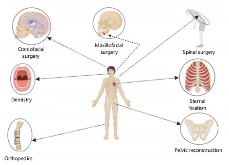
Versatility and expanding applications of titanium plates in surgical practices: Titanium plates, valued for their biocompatibility, strength, and corrosion resistance, are extensively used in various surgical fields. Beyond their established role in osteosynthesis, their versatility continues to grow in specialized procedures (Fig. 1).
In sternal fixation after open-heart surgery, titanium plates have been shown to provide superior stability compared to traditional sternal wiring, particularly in patients with morbid obesity. Their rigidity enhances fixation, reducing complications like sternal dehiscence and promoting better healing, leading to improved long-term outcomes46.
In cervical spine surgeries, titanium plates are preferred for stabilizing and fixing vertebrae due to their mechanical strength and ability to maintain structural integrity under load. Their use has been linked to improved fusion and alignment, facilitating better surgical and postoperative outcomes47.
Titanium plates are also crucial in craniofacial surgeries, particularly in orthognathic procedures for repositioning the upper maxilla. Custom-designed plates ensure precise bone repositioning, minimizing complications such as malocclusion and incorrect alignment, offering both functional and aesthetic improvements48.
|
|
The development of titanium alloy implants for mandibular defect reconstruction is another area of rapid advancement. These implants, tailored to individual anatomy, provide enhanced mechanical support, improved osseointegration, and a reduced risk of failure, contributing to successful functional and aesthetic outcomes4.
Ultimately, titanium’s unique properties make it indispensable across a wide range of surgical applications, from cardiovascular and spinal surgeries to craniofacial and mandibular reconstructions. As material science and surgical techniques advance, titanium plates will continue to expand their role in addressing complex clinical challenges, improving patient outcomes, and enhancing the precision of personalized surgical approaches.
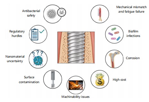
Challenges in the clinical application of titanium plate surfaces for osteogenesis: Despite remarkable advancements in titanium plate technologies, persistent challenges hinder their widespread clinical adoption in osteogenesis. These obstacles span mechanical, biological, and material science domains, necessitating multidisciplinary solutions to optimize implant performance and patient outcomes (Fig. 2).
Mechanical mismatch and long-term stability: Titanium is widely used in orthopedic and dental implants due to its excellent biocompatibility and mechanical properties. However, concerns regarding its long-term stability persist, especially in high-stress, load-bearing applications, where mechanical stresses, infection, and wear can lead to implant failure, loosening, or fracture over time3.
A critical limitation arises from the inherent mechanical disparity between titanium implants and native bone tissue. Titanium’s elastic modulus (110 GPa for Ti-6Al-4V) significantly exceeds that of cortical bone (10-30 GPa), leading to stress shielding a phenomenon where reduced mechanical loading on peri-implant bone triggers resorption and eventual implant loosening11. This mismatch is exacerbated in load-bearing applications such as spinal or joint replacements, where cyclic loading accelerates fatigue failure. Biomimetic designs that replicate bone’s anisotropic structure and modulus gradients are emerging as potential solutions, though scalability remains a hurdle2. Research into alternative alloys and surface modifications is needed to address this limitation.
Despite the promising potential of titanium implants with nanostructured surfaces to enhance osseointegration and offer antimicrobial benefits, their long-term effects remain poorly understood. More comprehensive studies are needed to evaluate the safety and durability of these nanomaterial-based modifications, ensuring that such innovations lead to sustained clinical improvements7.
Biofilm-associated infections and antimicrobial resistance: Infection control remains a major challenge in titanium implantology, despite advancements in antibacterial coatings. Implant-related infections, driven by biofilm-forming pathogens like Staphylococcus aureus and Pseudomonas aeruginosa, persist as a leading cause of failure. Biofilms are resistant to standard antibiotic treatments, complicating infection management and often leading to implant failure. They evade host immune responses and conventional antibiotics due to their extracellular polymeric matrix, complicating eradication. Preventing biofilm formation and ensuring sustained antibacterial efficacy remain critical challenges in titanium implant design1,37.
Although incorporating antibacterial materials into titanium surfaces shows promise, concerns over the safety and biocompatibility of certain agents persist. While antibacterial coatings (e.g., iodine-doped TiO2 nanotubes, silver nanoparticles) reduce initial bacterial adhesion, their efficacy diminishes over time. Regulatory concerns further limit clinical translation silver nanoparticles, though potent, risk cytotoxicity and inflammatory responses. Research must focus on both the antimicrobial effectiveness and the biocompatibility of these materials to ensure safe long-term use in implants1.
Another critical issue is the heat resistance of antibacterial coatings, particularly regarding sterilization. Titanium implants are commonly sterilized by autoclaving, which exposes them to high temperatures and pressures. Novel strategies, such as quorum-sensing inhibitors and immunomodulatory coatings, are under investigation to disrupt biofilm resilience without compromising biocompatibility. Coatings must retain their antibacterial properties after sterilization to ensure their ongoing effectiveness in clinical settings1.
Corrosion and ion release in physiological environments: Despite titanium’s corrosion resistance, localized degradation occurs in acidic microenvironments (e.g., inflammatory sites) or saline-rich tissues, releasing Ti4+ and Al3+ ions. Chronic exposure correlates with peri-implant fibrosis, osteolysis, and hypersensitivity reactions, particularly in patients with metabolic disorders. Recent case studies highlight ion accumulation in lymph nodes and distant organs, underscoring the need for real-time monitoring systems and corrosion-resistant alloys (e.g., Ti-Zr-Nb)49,50. The quest for more corrosion-resistant titanium alloys is ongoing. The Ti-Zr-Nb alloys are promising candidates due to their combination of improved biocompatibility54, enhanced mechanical properties52, and superior corrosion resistance49. Monitoring implant longevity, especially in patients with predisposing conditions, is essential
Long-term biocompatibility of nanomaterial modifications: Surface modification techniques are vital for enhancing the performance and biocompatibility of titanium implants in osteogenesis applications. However, they present several challenges. A major concern is surface contamination during treatment, which can undermine the desired outcomes such as improved osseointegration, antibacterial properties, or tissue compatibility. Contaminants from residual treatment by-products or environmental interactions can significantly affect implant performance, highlighting the need for strict process control to maintain integrity8.
Nanotopographical features (e.g., TiO2 nanotubes, nano-pillars) enhance early-stage osseointegration but raise concerns about long-term biological interactions. Animal models reveal nanoparticle migration into adjacent tissues, potentially inducing chronic inflammation or genotoxicity53-55. Furthermore, the degradation kinetics of bioactive coatings (e.g., hydroxyapatite-chitosan composites) remain poorly characterized in vivo, necessitating longitudinal studies to assess systemic impacts.
Additionally, the effectiveness of surface modifications is not uniform but depends on various factors, including the specific clinical context and patient variables. Factors such as age, bone quality, and underlying health conditions can influence how implants function post-surgery. As a result, personalized surface modification strategies are needed to address the diverse clinical scenarios encountered in orthopedic, maxillofacial, and spinal surgeries7.
Another limitation is the insufficient understanding of the long-term effects of surface treatments on implant stability and performance. While improvements in osseointegration and biocompatibility are often seen initially, the behavior of modified surfaces over time, under mechanical loading and in the biological environment, remains unclear. This knowledge gap is crucial for ensuring the long-term success of implants, especially in load-bearing applications8.
Machinability issues: Titanium alloys, essential in orthopedic and osteogenic applications, present significant machinability challenges due to their unique properties. Their low thermal conductivity, high chemical reactivity, and tendency to adhere to cutting tools complicate the machining process56. Titanium's inability to dissipate heat effectively leads to localized high temperatures, which can degrade both the material and cutting tools. The alloy’s reactivity with tool surfaces accelerates wear, contributing to potential tool failure.
Furthermore, machining titanium often results in poor surface finishes, which can hinder biocompatibility, osseointegration, and the effectiveness of surface treatments designed to promote bone growth. The high energy consumption required during machining also makes the process inefficient and costly. These challenges necessitate the development of advanced machining techniques and tool materials to optimize titanium processing.
Additive manufacturing (AM), particularly 3D printing, has introduced new complexities in machining titanium. While AM allows for customized and complex geometries, the microstructure and surface characteristics of AM-produced titanium differ from those of conventionally machined titanium. This variation requires tailored machining strategies and new approaches to overcome issues like tool wear and surface finish challenges.
To address these machinability issues, ongoing research is essential to explore novel machining technologies, advanced tool materials, and improved post-processing techniques. Such advancements could enhance the efficiency, quality, and cost-effectiveness of titanium implant production, leading to improved outcomes in osteogenesis and other medical applications.
Economic and logistical barriers: High manufacturing costs of titanium implants, driven by complex and energy-intensive processing and stringent quality control, present a major challenge in medical applications and limit accessibility in resource-constrained settings56. Additive manufacturing, though enabling patient-specific designs, requires costly post-processing to achieve optimal surface finishes. Additionally, regulatory pathways for novel coatings and technologies or 3D-printed implants demand extensive preclinical validation, delaying clinical adoption.
Moreover, titanium processing presents inherent challenges, such as rapid wear of machining tools and difficulties in achieving high-quality surface finishes, which increase operational costs. The high energy consumption required for processing titanium further complicates cost-efficiency, making it difficult for manufacturers to produce affordable, high-performance implants56.
Regulatory hurdles: The path from laboratory research to clinical application for new implant technologies, particularly in the field of titanium plate surface applications for osteogenesis, is often slow and fraught with complex regulatory hurdles.
Although titanium plates have demonstrated efficacy in certain surgeries, such as cervical spine procedures57. Introducing new implant designs or surface modifications into clinical practice involves stringent, time-consuming approval processes.
Regulatory approval is crucial, requiring rigorous safety, efficacy, and biocompatibility assessments. These barriers can delay promising innovations, especially those involving novel materials or surface treatments aimed at enhancing osseointegration or antibacterial properties. Furthermore, large-scale, multicenter clinical trials are essential to validate safety and long-term performance, but the high costs, logistical challenges, and lengthy follow-up periods complicate their execution. Regulatory bodies, such as the FDA or EMA, require extensive preclinical and clinical data before approving, and the process of demonstrating the safety and efficacy of new materials and designs can be a significant barrier to the timely introduction of these technologies into clinical practice38.
Clinical translation and personalized solutions: Patient-specific variables such as age, bone density, and comorbidities profoundly influence implant success. For example, osteoporotic patients exhibit delayed osseointegration due to reduced mesenchymal stem cell activity, while diabetic patients face higher infection risks. Current one-size-fits-all approaches inadequately address this diversity, highlighting the urgency for predictive biomarkers and AI-driven personalized implant designs.
Despite the significant and promising potential of titanium implant advancements with nanostructured surfaces to enhance osseointegration and offer antimicrobial benefits, their long-term effects remain poorly understood. Their clinical translation is impeded by regulatory delays, the necessity for extensive trials, and challenges in personalizing designs. More comprehensive studies are needed to evaluate the safety and durability of these nanomaterial-based modifications, ensuring that such innovations lead to sustained clinical improvements. Furthermore, initiatives to overcome these barriers will necessitate close collaboration among researchers, clinicians, and regulatory agencies to expedite the adoption of innovative technologies.
Prospects of titanium plate surface clinical application in osteogenesis: The future of titanium plate surface applications in osteogenesis is promising, with ongoing research and technological advancements paving the way for smarter, more effective, and patient-specific treatments.
Smart and customizable implants: Recent advances in titanium plate surface applications have paved the way for smart, customizable implants that could revolutionize osteogenesis. Smart implants, equipped with real-time sensors, allow continuous monitoring of implant stability, bone healing, and early infection detection, enabling early intervention and improved patient outcomes8. This shift toward proactive care allows for timely adjustments based on real-time data.
In parallel, 3D printing is transforming implant customization, offering personalized solutions that enhance osseointegration and long-term implant stability. These implants can be tailored in terms of shape, porosity, and surface characteristics to optimize bone growth and minimize complications2.
Drug-eluting implants also offer significant benefits, releasing controlled doses of therapeutic agents at the site of implantation to promote healing, reduce infection risk, and optimize tissue regeneration. For instance, localized antibiotic delivery can combat infection without systemic effects, while growth factor-releasing surfaces accelerate bone healing37.
Looking forward, Artificial Intelligence (AI) and machine learning (ML) are poised to further transform implant design by analyzing patient data to create anatomically precise, biologically tailored implants. These technologies can predict outcomes, identify complications, and personalize treatment plans, improving overall care2.
Thus, the integration of smart implants, 3D printing, drug-eluting technologies, and AI is set to redefine titanium implant therapy in osteogenesis, offering precision, efficiency, and patient-centered care.
Advanced surface modification technologies: Advancements in surface modification technologies are enhancing titanium plates for osteogenesis, particularly by promoting bone regeneration and improving implant integration. Innovations like nanomaterials, bioactive glasses, and growth factors are being incorporated to create bioactive surfaces that accelerate osseointegration and healing. Nanomaterials enhance surface area and cellular interactions, improving osteoblast adhesion and differentiation2. Bioactive glasses release ions to stimulate bone-forming cells, while growth factors like BMPs promote osteogenesis by encouraging mesenchymal stem cells to differentiate into osteoblasts.
Laser treatments are also gaining prominence, creating micro and nanostructures on titanium surfaces that mimic natural bone architecture, enhancing osseointegration and reducing bacterial adhesion8. When combined with coatings or functionalization, these modifications improve bioactivity and infection resistance, offering a robust strategy for implant success.
Surface functionalization with bioactive molecules, such as peptides or proteins, further enhances the biological response by promoting selective cell adhesion, proliferation, and osteogenesis. This method customizes implants to meet individual patient needs, optimizing bone formation at the implant-bone interface7.
Beyond techniques aimed solely at osteogenesis, broader advancements in biomaterial processing also offer valuable insights. For example, the technique of pulse electrodeposition, while explored for applying coatings to different biomaterials like zinc for biodegradable devices, highlights how precise control over processing parameters such as pulse frequency can significantly influence material properties, including microstructure, degradation rates, and crucially, biocompatibility aspects like blood compatibility58-60. Understanding these relationships, observed in materials like electrodeposited zinc, which showed benign hemolysis and cell compatibility with variations in blood compatibility depending on frequency, can inform the design of more effective titanium surface modifications58.
Collectively, the integration of nanomaterials, bioactive coatings, laser treatments, and surface functionalization offers transformative possibilities for titanium implants in osteogenesis. These technologies actively promote bone regeneration, improve implant stability, and reduce complications, marking a significant advancement in implant design for enhanced clinical outcomes.
Improved antibacterial strategies: Titanium implants in osteogenesis face ongoing challenges from implant-related infections, particularly biofilm formation. Recent advancements focus on enhancing antimicrobial properties without using harmful materials like heavy metals. Nano-structured surfaces have shown promise by increasing surface area and introducing physical or chemical cues that prevent bacterial adhesion and biofilm formation, providing a biocompatible, sustainable solution for clinical applications1,37.
Additionally, non-toxic antibacterial coatings using natural agents or synthetic peptides offer an effective, safe alternative to metal-based coatings. Bioactive materials, such as silver nanoparticles or antimicrobial polymers, provide long-term protection while maintaining biocompatibility, essential for seamless bone integration.
Immunomodulatory coatings, which regulate the immune response, further improve osteogenesis by reducing inflammation and enhancing cell recruitment to the implant site. These coatings also prevent biofilm formation and bolster the body's natural defenses, reducing post-surgical infections and improving clinical outcomes1,38.
The combination of nano-modifications, non-toxic coatings, and immunomodulatory features provides a comprehensive approach to infection prevention and bone regeneration. By targeting bacterial adhesion and supporting immune response, multi-functional implants promise to enhance implant resilience, reduce infection risk, and improve long-term osteogenic outcomes1.
Taken together, these innovative antibacterial strategies offer significant improvements in the safety and performance of titanium implants. By focusing on non-toxic, bioactive coatings, researchers are advancing toward implants that integrate better with bone tissue and provide robust infection protection, ultimately improving patient outcomes and reducing the need for invasive interventions.
Bioactive and biodegradable materials: The integration of bioactive and biodegradable materials into titanium plates marks a significant advancement in osteogenesis, balancing structural support with biological integration. Combining titanium with biodegradable materials allows implants to provide temporary mechanical stability while supporting tissue regeneration. These materials can also deliver growth factors and therapeutic agents, enhancing healing and reducing complications59.
Furthermore, hybrid systems that merge titanium’s strength with biologically favorable materials like bioactive ceramics, polymers, and natural biomolecules are gaining attention. These systems improve osseointegration, reduce implant failure risk, and better meet the physiological needs of bone healing45.
Magnesium alloys, which biodegrade and mimic bone properties, are also being explored for temporary implants. Their degradation products, such as magnesium ions, promote bone metabolism and mineralization, offering a physiological alternative to traditional materials60.
In addition to hybrid systems and biodegradable alloys, porous titanium structures, designed to mimic bone architecture, improve tissue ingrowth and osseointegration, enhancing implant stability and reducing complications. This approach accelerates healing by promoting vascularization and bone cell growth11.
After all, bioactive and biodegradable titanium implants, including hybrid systems, biodegradable alloys, and porous structures, offer promising solutions for bone regeneration. These advancements enhance tissue integration, improve patient outcomes, and reduce complications in osteogenesis applications.
Enhanced diagnostic and monitoring techniques: Advancements in diagnostic and monitoring techniques are crucial for optimizing the clinical success of titanium implants in osteogenesis. High-resolution imaging, particularly micro-computed tomography (micro-CT), enables precise 3D visualization of the bone-implant interface at micro and nanoscale levels. This provides valuable insights into osseointegration, implant positioning, and material integration, supporting the development of improved implant designs and surface modifications2.
Non-invasive monitoring methods, including in vivo fluorescence imaging, strain gauges, and biomechanical systems, offer continuous, real-time assessments of implant stability and bone healing. These technologies help detect early complications such as implant loosening or infection, allowing for timely interventions that improve patient outcomes and reduce the risk of implant failure8.
Together, these innovations are revolutionizing implant technology, facilitating personalized, data-driven patient care, and paving the way for advancements in regenerative medicine.
Refined surgical planning using CAD/CAM: The integration of Computer-Assisted Design and Manufacturing (CAD/CAM) technologies has revolutionized the customization of titanium implants for osteogenesis, enhancing surgical planning and precision61,62.The CAD/CAM allows for the design of titanium plates tailored to individual anatomical structures using patient-specific imaging data (CT, MRI), ensuring a precise fit and optimizing mechanical properties, which in turn improves osseointegration and reduces complications63.
Moreover, CAD/CAM enables virtual simulation of surgeries, allowing surgeons to test implant positioning and anticipate challenges before the procedure. This preoperative planning improves clinical outcomes, reduces surgical risks, and shortens implant manufacturing time, ultimately lowering costs.
Advancements in CAD/CAM, particularly 3D printing and rapid prototyping, further enhance implant precision, enabling the creation of complex designs that were previously unattainable. These innovations contribute to personalized, patient-centered care, resulting in better outcomes and faster recovery64,65.
Briefly, CAD/CAM technologies are transforming titanium implant fabrication, advancing both the precision and efficacy of osteogenesis surgeries and marking a significant step toward personalized regenerative medicine.
Integration of stem cells: The integration of mesenchymal stem cells (MSCs) with titanium implants has emerged as a promising strategy to enhance bone regeneration and osseointegration. The MSCs, known for their self-renewal, multi-lineage differentiation, and paracrine signaling, can accelerate bone healing and strengthen the bond between the implant and surrounding tissue66. The MSCs derived from bone marrow, adipose tissue, and other sources have been widely studied for their osteogenic potential, promoting the differentiation of osteoblasts and the formation of a supportive extracellular matrix8.
When combined with titanium plates, MSCs enhance osseointegration by promoting vascularization, reducing inflammation, and modulating the immune response to prevent infection and implant rejection. This combination also offers advantages over traditional grafting, facilitating faster recovery and improving implant stability. Recent research highlights the role of bioactive factors released by MSCs in supporting long-term implant function.
The clinical application of MSCs in titanium implant therapies has shown promise in treating non-union fractures, critical-size bone defects, and skeletal disorders. Advancements in tissue engineering have led to the development of composite implants with surface-modified titanium plates that provide an optimal microenvironment for MSC adhesion and differentiation.
Despite these advances, challenges remain, including optimizing stem cell sourcing, controlled delivery, and ensuring long-term clinical success. Issues related to immune rejection, tumorigenesis, and ethical concerns must also be addressed. Nonetheless, MSC integration with titanium implants holds significant potential for advancing bone regeneration therapies and improving patient outcomes in orthopedic and maxillofacial surgeries.
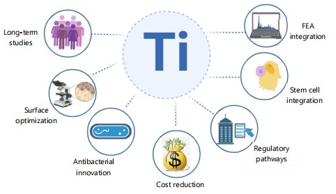
To optimize the clinical application of titanium implants for osteogenesis, a comprehensive set of strategic interventions is recommended to enhance efficacy, accessibility, and long term success (Fig. 3).
Long-term outcomes research: To optimize the clinical application of titanium implants for osteogenesis, it is imperative to conduct comprehensive long-term clinical studies that evaluate the stability, mechanical integrity, corrosion resistance, and wear characteristics of these implants, especially those with advanced surface modifications. Such studies should encompass diverse patient populations, including individuals with comorbidities, to ensure the generalizability of findings2.
|
The ongoing quest for more corrosion-resistant titanium alloys has brought significant attention from researchers. The Ti-Zr-Nb alloys are promising candidates due to their improved biocompatibility, enhanced mechanical properties, and superior corrosion resistance. Compared to aluminum and vanadium, present in the widely used Ti-6Al-4V alloy, niobium and zirconium are generally considered more biocompatible51. The Ti-Zr-Nb alloys can be designed to have a lower elastic modulus, which is closer to that of bone, reducing stress shielding and promoting better bone ingrowth52. Moreover, alloying with Zr and Nb can improve the passive layer stability on titanium, reducing ion release49.
For instance, a 10-year retrospective study involving 511 sandblasted, large-grit, acid-etched (SLA) implants in 303 partially edentulous patients reported a success rate of 97.0% and a survival rate of 98.8%, with a peri-implantitis rate as low as 1.8%67. Additionally, a systematic review highlighted that bioactive surface modifications, such as collagen-based coatings, significantly enhance osseointegration and prolong the lifespan of dental implants68. Furthermore, a scoping review emphasized that implant surface properties, including topography and energy, can influence epigenetic modifications, thereby affecting osseointegration and peri-implant health69. These findings underscore the necessity of long-term, multicentric clinical trials to validate the efficacy and safety of surface-modified titanium implants across varied patient demographics.
Optimize surface modifications: Improving the clinical performance of titanium implants requires that surface modification strategies be refined to simultaneously enhance osseointegration and reduce infection risks. To this end, we recommend prioritizing the development of advanced bioactive coatings that incorporate osteoinductive and immunomodulatory agents into hierarchically structured surfaces11. For example, engineering nanostructured and micro roughened topographies via methods such as microarc oxidation, laser ablation, or anodization has been shown to promote osteoblast adhesion, differentiation, and robust implant–bone integration while also deterring bacterial colonization70,71. In parallel, integrating controlled-release systems for growth factors (e.g., BMP-2) or antibacterial ions (e.g., Ag+, Mg2+, Sr2+) within these coatings can provide a synergistic effect that both stimulates bone regeneration and mitigates local infection71. Furthermore, advanced fabrication techniques such as layer-by-layer assembly and additive manufacturing enable precise tailoring of surface chemistry and topography, thereby ensuring reproducible and durable bioactivity that bridges the gap between laboratory success and clinical translation. Ultimately, these multi-modal surface modifications are expected to improve implant fixation, reduce revision rates, and enhance patient outcomes.
Antibacterial innovations: Developing safe, biocompatible antibacterial coatings is imperative for preventing implant associated infections a persistent challenge in orthopedic and dental surgeries. Recent advances in nanotechnology and biomaterial engineering have enabled the creation of multifunctional coatings that not only deter bacterial adhesion and disrupt biofilm formation but also actively modulate local immune responses to promote healing and osseointegration. Strategies include the incorporation of antimicrobial metal ions (e.g., silver, copper, and zinc), antimicrobial peptides, and stimuli responsive polymers designed for on demand release of bactericidal agents in response to infection cues1. These “smart” coatings target the early stages of bacterial colonization by repelling pathogens and, when needed, releasing therapeutic agents locally to minimize systemic side effects and reduce the risk of antibiotic resistance. Moreover, the integration of nanoscale surface topographies with biochemical functionalities has shown promise in enhancing osteogenic cell adhesion while suppressing bacterial growth, thereby fostering a microenvironment that supports tissue regeneration and long term implant stability. Collectively, these innovations hold significant potential to improve clinical outcomes by reducing revision surgeries and enhancing overall implant performance70,72,73.
Cost reduction and accessibility: Reducing production costs while upholding rigorous quality standards is pivotal for the broad clinical dissemination of titanium implants. Recent advances in additive manufacturing have demonstrated that streamlined fabrication processes can significantly lower costs without compromising implant integrity or biocompatibility56. In parallel, innovative surface modification strategies such as micro-arc oxidation, plasma spraying, and the deposition of bioactive coatings have been shown to improve osteointegration and reduce the need for costly post processing treatments74-77. Together, these approaches offer a promising pathway to not only enhance the clinical performance of titanium implants but also to increase their accessibility, particularly in low- and middle-income regions. Continued research into the scalability and long-term performance of these cost-effective modifications is essential to facilitate their translation from bench to bedside, ultimately supporting sustainable, high-quality healthcare worldwide.
3D printing and customization: Recent advances in additive manufacturing have enabled the fabrication of patient specific titanium implants with finely controlled microstructures and porosities that mimic natural bone architecture. These customized implants offer improved mechanical compatibility and significantly enhance osteointegration by promoting cell attachment, proliferation, and differentiation at the implant–bone interface2. Tailoring implant geometry and surface topography using 3D printing not only optimizes load transfer and reduces stress shielding but also enables the incorporation of bioactive coatings and drug delivery systems, thereby improving clinical outcomes in complex reconstructive procedures78,79. Clinical studies have demonstrated that such personalized implants lead to faster osseointegration and higher patient satisfaction in applications ranging from craniofacial reconstructions to orthopedic tumor resection80,81. As 3D printing technology continues to evolve, its role in producing customized implants with enhanced osteogenic capabilities is poised to transform patient-specific regenerative strategies and improve long-term implant success.
Multidisciplinary collaboration: Advancing titanium implant surface modifications for clinical osteogenesis mandates active, long-term collaboration between materials scientists, biomedical engineers, and clinicians. Such interdisciplinary efforts are essential to integrate novel insights from nanotechnology, bioactive coatings, and laser-based surface engineering into implant design1. The synthesis of expertise across these fields not only accelerates the translation of bench-top innovations into improved clinical protocols but also fosters the standardization of implant surface characterization and evaluation methodologies. Recent studies have demonstrated that combining laser ablation techniques with biofunctional coatings significantly enhances both osteogenic and antibacterial properties82,83. Furthermore, clinical evaluations indicate that multidisciplinary research can reduce implant failure rates by optimizing surface parameters tailored to patient-specific bone physiology84. Future research should aim to establish integrated platforms that enable iterative feedback between laboratory findings and clinical outcomes, ensuring that titanium implant designs not only achieve rapid osseointegration but also demonstrate long-term stability and resistance to peri-implant infections.
Standardize testing and evaluation: To advance the clinical translation of titanium implants for osteogenesis, it is imperative to adopt standardized testing protocols that encompass comprehensive mechanical, biological, and immunological evaluations. Uniform methodologies-ranging from detailed mechanical assessments (e.g., fatigue, tensile strength, and fracture resistance) and precise surface characterizations (including quantification of roughness, hydrophilicity, and chemical composition) to rigorous in vitro/in vivo assays simulating the clinical environment are essential for generating reproducible and predictive data7. In parallel, well designed clinical trials using standardized outcome measures, such as implant stability quotient (ISQ), marginal bone level changes, and histomorphometric analyses, are critical to validate implant safety and long term efficacy across diverse patient populations. Moreover, harmonizing evaluation methodologies with internationally recognized standards, such as ISO 10993-1:2018 for biological safety and ASTM guidelines for mechanical performance (https://www.iso.org/standard/68936.html)-will ensure reproducibility and facilitate cross-study comparability. Advances in analytical imaging and quantitative techniques now enable a more robust appraisal of implant performance, ultimately facilitating improved reproducibility and inter-study comparability in both preclinical and clinical settings85,86.
Use finite element analysis: Finite element analysis (FEA) should be integrated into the design phase of titanium implants to optimize mechanical properties and predict in vivo performance under physiological loading. By simulating stress distribution, micromotion, and fatigue behavior, FEA enables designers to refine implant geometry, surface modifications, and thread designs to minimize stress concentrations and mitigate the risk of implant failure45,47. Recent investigations have demonstrated that FEA-driven optimization can enhance osseointegration by promoting favorable load transfer at the bone-implant interface87,88. Moreover, FEA supports the development of patient-specific implants that account for variations in bone quality and anatomical geometry, ultimately improving long-term clinical outcomes89. The inhe integration of these computational tools into implant design not only improves the implant’s strength and reliability but also fosters the translation of biomechanical principles into clinical practice for enhanced osteogenesis.
Enhance machining technologies: Advancements in machining technologies for titanium alloys-including improvements in tool design, advanced cooling strategies, and the integration of additive manufacturing processes are critical to refining the precision and reducing the cost of implant production56. Optimizing these processes minimizes material wastage, enhances surface integrity, and supports the fabrication of patient-specific implants with superior biomechanical performance. Such enhancements not only ensure consistent quality and reproducibility but also expand the accessibility of high-quality titanium implants for clinical osteogenesis applications. Recent studies have demonstrated that the synergistic use of optimized machining parameters and additive manufacturing techniques can significantly improve both the microstructural characteristics and the osseointegration potential of titanium implants90.
Improve regulatory processes: Optimizing the regulatory framework is critical to accelerating the translation of innovative titanium implant surface modifications into clinical practice. Streamlined, transparent, and harmonized review pathways not only shorten approval timelines for new materials but also safeguard patient safety by integrating robust post-market surveillance and real world evidence into the evaluation process7. Recent advances in regulatory science, such as adaptive review frameworks and the incorporation of real-world data, have demonstrated that aligning regulatory practices across regions can reduce development costs and improve patient access without compromising safety91. Moreover, enhanced collaboration among regulatory agencies, manufacturers, and clinicians is essential to establish clear, flexible guidelines that can readily accommodate emerging technologies in implant design92. Together, these measures will foster the timely integration of innovative surface modifications, ultimately enhancing the efficacy, accessibility, and long-term clinical success of titanium implants in osteogenesis.
Focus on regenerative medicine: Advancing regenerative medicine is crucial for the next generation of titanium implant therapies. In particular, integrating mesenchymal stem cells (MSCs) with titanium implants has been shown to promote osteogenic differentiation, enhance matrix mineralization, and modulate the local inflammatory environment-thereby accelerating bone healing and improving osseointegration66. Preclinical investigations have demonstrated that MSC based interventions can substantially increase new bone formation and implant stability in both healthy and compromised bone conditions93,94. Continued research into optimized cell sourcing, scaffold design, and growth factor delivery will likely revolutionize clinical outcomes, offering patient tailored solutions for complex bone injuries95,96. Implementing these regenerative medicine approaches is anticipated to significantly enhance the clinical performance of titanium implants, thereby solidifying their role in modern orthopedic and reconstructive surgery.
Finally, by adopting these recommendations, significant advancements in titanium implant technologies for osteogenesis can be realized. This progress holds the potential to enhance clinical outcomes and reinforce the pivotal role of titanium implants in contemporary orthopedic and reconstructive surgery.
CONCLUSION
Titanium plate surfaces have emerged as a transformative innovation in the field of osteogenesis, playing a pivotal role in advancing bone regeneration and implantation technologies. This comprehensive review underscores the remarkable progress achieved in surface modification techniques, antibacterial innovations, and the development of personalized implant designs, all of which have collectively contributed to enhanced clinical outcomes and patient satisfaction. Despite these advancements, significant challenges persist, including concerns related to long-term implant stability, cost-effectiveness, infection prevention, and the seamless translation of cutting-edge technologies from laboratory research to clinical practice. Looking ahead, the future of titanium plate applications in osteogenesis appears exceptionally promising, driven by the advent of smart implants, next-generation surface modification strategies, refined antibacterial solutions, and the integration of regenerative medicine principles. By addressing these challenges through sustained innovation, robust multidisciplinary collaborations, and a patient-centered approach, the field is poised to not only improve the quality of life for countless patients but also to redefine the boundaries of bone regeneration and tissue engineering. This evolution holds the potential to unlock novel therapeutic avenues and establish new benchmarks in musculoskeletal healthcare.
SIGNIFICANCE STATEMENT
Titanium plate surfaces have revolutionized osteogenesis by advancing bone regeneration and implant technologies. This review outlines major progress in surface modifications, antibacterial strategies, and personalized implant designs, leading to improved clinical outcomes. Despite these achievements, challenges such as long-term stability, cost-effectiveness, and clinical translation remain. Future developments, including smart implants and regenerative medicine integration, promise to enhance patient care and redefine standards in musculoskeletal healthcare.
REFERENCES
- Lee, J.W., J.A. Cho, Y.J. Roh, M.A. Han and J.U. Jeong et al., 2024. Antibacterial immunonegative coating with biocompatible materials on a nanostructured titanium plate for orthopedic bone fracture surgery. Biomater. Res., 28.
- Dobrzyński, M., M. Szymonowicz, J. Nowicka, M. Pajączkowska and A. Nikodem et al., 2024. Three-dimensional-printed modular titanium alloy plates for osteosynthesis of the jawbone. Biomedicines, 12.
- Zhou, W., T. Wang, Y. Gan, J. Yang and H. Zhu et al., 2020. Effect of micropore/microsphere topography and a silicon-incorporating modified titanium plate surface on the adhesion and osteogenic differentiation of BMSCs. Artif. Cells Nanomed. Biotechnol., 48: 230-241.
- Lin, H., X. Zhong, N. Zhou and X.P. Huang, 2023. Mandibular reconstruction after excision of recurrent odontogenic keratocyst using a novel mandibular distraction osteogenesis method-A case report. Head Face Med., 19.
- Yin, W., R.B.N. Raby, Y. Li, Z. Li, M. Sun and Z. Huang, 2023. An alternating magnetic field-controlled drug delivery system based on 4,4′-azobis (4-cyanovaleric acid)-functioned Fe3O4@chitosan nanoparticles. Bioengineering, 10.
- Yang, Y., X. Wang, R.J. Miron and X. Zhang, 2019. The interactions of dendritic cells with osteoblasts on titanium surfaces: An in vitro investigation. Clin. Oral Invest., 23: 4133-4143.
- Li, H., J. Huang, Y. Wang, Z. Chen and X. Li et al., 2022. Nanoscale modification of titanium implants improves behaviors of bone mesenchymal stem cells and osteogenesis in vivo. Oxid. Med. Cell. Longevity, 2022.
- Chen, Y.W., T. Chiang, I.H. Chen, D.Y. Yuh, H.Y. Tseng, C.W. Wang and H.H. Hou, 2024. Titanium surfaces with a laser-produced microchannel structure enhance pre-osteoblast proliferation, maturation, and extracellular mineralization in vitro. Int. J. Mol. Sci., 25.
- Liu, X., S. Chen, H. Ding and J.K.H. Tsoi, 2024. Antifungal ability of novel silane on titanium implant surface. Coatings, 14.
- Matsuura, T., K. Komatsu, T. Suzumura, S. Stavrou, M.L. Juanatas, W. Park and T. Ogawa, 2024. Enhanced functionality and migration of human gingival fibroblasts on vacuum ultraviolet light-treated titanium: An implication for mitigating cellular stress to improve peri-implant cellular reaction. J. Prosthodontic Res., 69: 249-258.
- Abd-Elaziem, W., M.M. Mohammed, H.M. Yehia, T.A. Sebaey and T. Khan, 2024. Porous titanium for medical implants. MultiDiscip. Mater. Chronicles., 1: 1-18.
- Wu, B., Y. Tang, K. Wang, X. Zhou and L. Xiang, 2022. Nanostructured titanium implant surface facilitating osseointegration from protein adsorption to osteogenesis: The example of TiO2 NTAs. Int. J. Nanomed., 17: 1865-1879.
- Anitua, E., L. Piñas, A. Murias, R. Prado and R. Tejero, 2015. Effects of calcium ions on titanium surfaces for bone regeneration. Colloids Surf. B: Biointerfaces., 130: 173-181.
- Wehner, C., M. Laky, H.A. Shokoohi-Tabrizi, C. Behm, A. Moritz, X. Rausch-Fan and O. Andrukhov, 2021. Effects of Er:YAG laser irradiation of different titanium surfaces on osteoblast response. J. Mater. Sci: Mater. Med., 32.
- Zhang, T., X. Zhang, M. Mao, J. Li, T. Wei and H. Sun, 2020. Chitosan/hydroxyapatite composite coatings on porous Ti6Al4V titanium implants: In vitro and in vivo studies. J. Periodontal Implant Sci., 50: 392-405.
- Fraioli, R., P.M. Tsimbouri, L.E. Fisher, A.H. Nobbs and B. Su et al., 2017. Towards the cell-instructive bactericidal substrate: Exploring the combination of nanotopographical features and integrin selective synthetic ligands. Sci. Rep., 7.
- Lotz, E.M., M.B. Berger, B.D. Boyan and Z. Schwartz, 2020. Regulation of mesenchymal stem cell differentiation on microstructured titanium surfaces by semaphorin 3A. Bone, 134.
- Deng, J., D.J. Cohen, M.B. Berger, E.L. Sabalewski, M.J. McClure, B.D. Boyan and Z. Schwartz, 2023. Osseointegration of titanium implants in a botox-induced muscle paralysis rat model Is sensitive to surface topography and semaphorin 3A treatment. Biomimetics, 8.
- Song, A., F. Jiang, Y. Wang, M. Wang and Y. Wu et al., 2022. Semaphorin3A promotes osseointegration of titanium implants in osteoporotic rabbits. Clin. Oral Invest., 26: 969-979.
- Kirschneck, C., F. Wolf, F. Cieplik, M. Blanck-Lubarsch, P. Proff and A. Schröder, 2020. Impact of NSAID etoricoxib on side effects of orthodontic tooth movement. Ann. Anat. Anatomischer Anz., 232.
- Virdi, A.S., J. Irish, K. Sena, M. Liu, H.Z. Ke, M.A. McNulty and D.R. Sumner, 2015. Sclerostin antibody treatment improves implant fixation in a model of severe osteoporosis. J. Bone Joint Surg., 97: 133-140.
- Cheng, H., W. Xiong, Z. Fang, H. Guan and W. Wu et al., 2016. Strontium (Sr) and silver (Ag) loaded nanotubular structures with combined osteoinductive and antimicrobial activities. Acta Biomater., 31: 388-400.
- Susin, C., M. Qahash, G. Polimeni, P.H. Lu and H.S. Prasad et al., 2010. Alveolar ridge augmentation using implants coated with recombinant human bone morphogenetic protein-7 (rhBMP-7/rhOP-1): Histological observations. J. Clin. Periodontology, 37: 574-581.
- García-Gareta, E., J. Hua, A. Orera, N. Kohli, J.C. Knowles and G.W. Blunn, 2017. Biomimetic surface functionalization of clinically relevant metals used as orthopaedic and dental implants. Biomed. Mater., 13.
- Kuroda, S., R. Wazen, P. Moffatt, E. Tanaka and A. Nanci, 2013. Mechanical stress induces bone formation in the maxillary sinus in a short-term mouse model. Clin. Oral Invest., 17: 131-137.
- Martinez‐Marquez, D., K. Terhaer, P. Scheinemann, A. Mirnajafizadeh, C.P. Carty and R.A. Stewart, 2020. Quality by design for industry translation: Three-dimensional risk assessment failure mode, effects, and criticality analysis for additively manufactured patient-specific implants. Eng. Rep., 2.
- Schliephake, H., J. Rublack, N. Aeckerle, A. Förster, B. Schwenzer, J. Reichert and D. Scharnweber, 2015. In vivo effect of immobilisation of bone morphogenic protein 2 on titanium implants through nano-anchored oligonucleotides. Eur. Cells Mater., 30: 28-40.
- Huang, Z., Y. Li, W. Yin, R.B.N. Raby, H. Liang and B. Yu, 2025. A magnetic-guided nano-antibacterial platform for alternating magnetic field controlled vancomycin release in Staphylococcus aureus biofilm eradication. Drug Delivery Transl. Res., 15: 1249-1264.
- Huang, Z., R.B.N. Raby, W. Yin, M. Zhang and Z. Li, 2025. Development of ROS-responsive collagen-based hemostatic sponges for the repair of MRSA-infected wounds. Int. J. Biol. Macromol., 305.
- Haugen, H.J., S. Makhtari, S. Ahmadi and B. Hussain, 2022. The antibacterial and cytotoxic effects of silver nanoparticles coated titanium implants: A narrative review. Materials, 15.
- Yang, M., S. Qiu, E. Coy, S. Li and K. Załęski et al., 2022. NIR-responsive TiO2 biometasurfaces: Toward in situ photodynamic antibacterial therapy for biomedical implants. Adv. Mater., 34.
- Kalelkar, P.P., M. Riddick and A.J. García, 2022. Biomaterial-based antimicrobial therapies for the treatment of bacterial infections. Nat. Rev. Mater., 7: 39-54.
- Lei, Z., H. Liang, W. Sun, Y. Chen, Z. Huang and B. Yu, 2024. A biodegradable PVA coating constructed on the surface of the implant for preventing bacterial colonization and biofilm formation. J. Orthop. Surg. Res., 19.
- Wu, Y., H. Tang, L. Liu, Q. He and L. Zhao et al., 2020. Biomimetic titanium implant coated with extracellular matrix enhances and accelerates osteogenesis. Nanomedicine, 15: 1779-1793.
- Soro, N., E.G. Brodie, A. Abdal-hay, A.Q. Alali, D. Kent and M.S. Dargusch, 2022. Additive manufacturing of biomimetic titanium-tantalum lattices for biomedical implant applications. Mater. Des., 218.
- Oltean-Dan, D., G.B. Dogaru, E.M. Jianu, S. Riga and M. Tomoaia-Cotisel et al., 2021. Biomimetic composite coatings for activation of titanium implant surfaces: Methodological approach and in vivo enhanced osseointegration. Micromachines, 12.
- Yang, X., N.F. Chen, X.L. Huang, S. Lin, Q.Q. Chen, W.M. Wang and J.S. Chen, 2023. Iodine-doped TiO2 nanotube coatings: A technique for enhancing the antimicrobial properties of titanium surfaces against Staphylococcus aureus. J. Orthop. Surg. Res., 18.
- Zhang, H., Y. Yuan, H. Xue, R. Yu, X. Jin, X. Wu and H. Huang, 2023. Reprogramming mitochondrial metabolism of macrophages by miRNA-released microporous coatings to prevent peri-implantitis. J. Nanobiotechnol., 21.
- Zhao, C., W. Liu, M. Zhu, C. Wu and Y. Zhu, 2022. Bioceramic-based scaffolds with antibacterial function for bone tissue engineering: A review. Bioact. Mater., 18: 383-398.
- Teodorescu, M., M. Bercea and S. Morariu, 2019. Biomaterials of PVA and PVP in medical and pharmaceutical applications: Perspectives and challenges. Biotechnol. Adv., 37: 109-131.
- Chen, T., C. Brial, M. McCarthy, R.F. Warren and S.A. Maher, 2021. Synthetic PVA osteochondral implants for the knee joint: Mechanical characteristics during simulated gait. Am. J. Sports Med., 49: 2933-2941.
- Chen, Y., J. Song, S. Wang and W. Liu, 2021. PVA-based hydrogels: Promising candidates for articular cartilage repair. Macromol. Biosci., 21.
- Liu, F.J., Y.C. Liu, Y.C. Hu, X.K. Ding and J. Miao, 2023. Novel three-holed titanium plate fixation during open door laminoplasty for cervical spondylotic myelopathy: Comparison with conventional titanium plate. Orthop. Surg., 15: 197-204.
- Wang, W., X. Cai, X. Liu, G. Wang, H. Kang and S. Qian, 2024. Special contoured pelvic brim reconstruction titanium plate combined with trans-plate buttress screws (quadrilateral screws) for acetabular fractures with quadrilateral plate involvement through the anterior ilioinguinal approach. Front. Surg., 11.
- Fang, S., L. Zhang, Y. Yang, Y. Wang, J. Guo and L. Mi, 2023. Finite element analysis comparison of type 42A2 fracture fixed with external titanium alloy locking plate and traditional external fixation frame. J. Orthop. Surg. Res., 18.
- Atik, C. and D. Atik, 2023. The results of rigid titanium plate reinforcement and only conventional wire methods in sternal fixation in morbidly obese patients. Braz. J. Cardiovasc. Surg., 38.
- Xu, H., J. Wu, H. Xie, W. Wen, H. Xu, J. Du and J. Miao, 2022. Biomechanical behaviour of tension-band-reconstruction titanium plate in open-door laminoplasty: A study based on finite element analysis. BMC Musculoskeletal Disord., 23. .
- Wu, S., W. Zhang and J. Shi, 2024. Evaluation of the efficacy of three times titanium plate gradual fixation method in the treatment of extracapsular condylar fractures. J. Craniofacial Surg., 35: 1289-1291.
- Zhang, S., X. Du, W. Shi and S. Xiang, 2024. Effect of low dissolved oxygen concentration on the tribocorrosion properties and ion release of Ti-13Nb-13Zr. Corros. Eng. Sci. Technol., 59: 236-248.
- Fokter, S.K., Ž. Ledinek, M.K. Dujić and I. Novak, 2024. Extreme serum titanium concentration induced by acetabular cup failure: Unveiling a unique scenario of titanium alloy debris accumulation. Bioengineering, 11.
- Hassan, T.H. and N.A. Kalluvalappil, 2024. Allergic reaction of poly-ether-ether-ketone versus titanium implants: A posttest-only control group design experimental study using a rabbit model. Clin. Implant Dent. Relat. Res., 26: 671-678.
- Wang, X., Y. Gao, M. Bao, Y. Liu, L. Yang and E. Zhang, 2024. Enhancing the in vitro tribocorrosion resistance of titanium alloy by precipitation of nano-Ti2Cu phases. J. Mater. Eng. Perform., 33: 12966-12975.
- Mohamed, H.R.H., 2015. Estimation of TiO2 nanoparticle-induced genotoxicity persistence and possible chronic gastritis-induction in mice. Food Chem. Toxico., 83: 76-83.
- Huang, S., F. Guo, N. Liu, K. Hu and C. Liu, 2024. Cellular experimental study and mechanism of surface modification of 3D printed titanium materials to modulate the sealing and antimicrobial properties of oral implants. Eur. J. Inflammation, 22.
- Sreenivasagan, S., A.K. Subramanian, K.G. Mohanraj and R.S. Kumar, 2023. Assessment of toxicity of green synthesized silver nanoparticle-coated titanium mini-implants with uncoated mini-implants: Comparison in an animal model study. J. Contemp. Dent. Pract., 24: 944-950.
- Polishetty, A., G. Bolar, J. Nomani and G. Littelfair, 2024. Evaluating productivity characteristics of laser engineered net shaping titanium alloy. Int. J. Adv. Manuf. Technol., 135: 1377-1383.
- Gómez, S.G., M.P. Ginebra, F.J. Gil, R.I. Barraquer and J.M. Manero, 2024. Antibacterial and cytocompatible silver coating for titanium Boston keratoprosthesis. Front. Bioeng. Biotechnol., 12.
- Wu, S., Y. Luo, W. Hu, Y. Chen and Z. Huang, 2022. Effect of pulse frequency on the microstructure and the degradation of pulse electroformed zinc for fabricating the shell of biodegradable dosing pump. Bioengineering, 9.
- Kumar, C.M.P., A. Lakshmikanthan, M.P.G. Chandrashekarappa, D.Y. Pimenov and K. Giasin, 2021. Electrodeposition based preparation of Zn-Ni alloy and Zn-Ni-WC nano-composite coatings for corrosion-resistant applications. Coatings, 11.
- Kumar, C.M.P., M.P.G. Chandrashekarappa, R.M. Kulkarni, D.Y. Pimenov and K. Giasin, 2021. The effect of Zn and Zn-WO3 composites nano-coatings deposition on hardness and corrosion resistance in steel substrate. Materials, 14.
- Hu, M., M. Li, R. Ma, X. Li and X. Ren et al., 2024. Biomechanical analysis of titanium-alloy and biodegradable implants in dual plate osteosynthesis for AO/ASIF type 33-C2 fractures. Heliyon, 10.
- Shang, Y., Y. Bi, Y. Cao and Y. Wang, 2023. Finite element analysis of titanium anatomic plate and titanium reconstructive plate for treatment of extra-articular fractures of the scapula. J. Orthop. Surg. Res., 18.
- Sun, Y., W. Du, C. Xu, Y. Lin, X. Liu and E. Luo, 2020. Applications of computer-aided design/manufacturing technology in treatment of hemifacial microsomia. J. Craniofacial Surg., 31: 1133-1136.
- del Castillo, R. and J. Ata-Ali, 2023. The clinical use of computer aided designed/computer aided manufactured titanium nitride coated implant abutments: Surgical and prosthetic considerations-A case series. J. Esthetic Restor. Dent., 35: 1008-1021.
- Verschueren, J., D.E. Meuffels, E.E. Bron, S. Klein and G.J. Kleinrensink et al., 2018. Possibility of quantitative T2-mapping MRI of cartilage near metal in high tibial osteotomy: A human cadaver study. J. Orthop. Res., 36: 1206-1212.
- Wang, J., P. Wu, H.L. Liu, L. Zhang, L.P. Liu, C.F. Ma and J.H. Chen, 2022. Polyetheretherketone versus titanium CAD-CAM framework for implant-supported fixed complete dentures: A retrospective study with up to 5-year follow-up. J. Prosthodontic Res., 66: 279-287.
- Donker, V.J.J., G.M. Raghoebar, C. Jensen‐Louwerse, A. Vissink and H.J.A. Meijer, 2022. Monolithic zirconia single tooth implant-supported restorations with CAD/CAM titanium abutments in the posterior region: A 1-year prospective case series study. Clin. Implant Dent. Relat. Res., 24: 125-132.
- Cheng, L., Y. Li, Q. Xia, M. Meng and Z. Ye et al., 2021. Enamel matrix derivative (EMD) enhances the osteogenic differentiation of bone marrow mesenchymal stem cells (BMSCs). Bioengineered, 12: 7033-7045.
- Smeets, R., B. Stadlinger, F. Schwarz, B. Beck-Broichsitter and O. Jung et al., 2016. Impact of dental implant surface modifications on osseointegration. BioMed Res. Int., 2016.
- Al Shayeb, M., S. Elfadil, H. Abutayyem, A. Shqaidef, M.M. Marrapodi, M. Cicciù and G. Minervini, 2024. Bioactive surface modifications on dental implants: A systematic review and meta-analysis of osseointegration and longevity. Clin. Oral Invest., 28.
- Kunrath, M.F., C. Garaicoa-Pazmino, P.M. Giraldo-Osorno, A.H. Mustafa, C. Dahlin, L. Larsson and F. Asa'ad, 2024. Implant surface modifications and their impact on osseointegration and peri-implant diseases through epigenetic changes: A scoping review. J. Periodontal Res., 59: 1095-1114.
- Lu, X., Z. Wu, K. Xu, X. Wang and S. Wang et al., 2021. Multifunctional coatings of titanium implants toward promoting osseointegration and preventing infection: Recent developments. Front. Bioeng. Biotechnol., 9.
- Zhu, G., G. Wang and J.J. Li, 2021. Advances in implant surface modifications to improve osseointegration. Mater. Adv., 2: 6901-6927.
- Akshaya, S., P.K. Rowlo, A. Dukle and A.J. Nathanael, 2022. Antibacterial coatings for titanium implants: Recent trends and future perspectives. Antibiotics, 11.
- Wang, H., C. Xiong, Z. Yu, J. Zhang, Y. Huang and X. Zhou, 2022. Research progress on antibacterial coatings for preventing implant-related infection in fractures: A literature review. Coatings, 12.
- Yang, S.P., T.M. Lee and T.S. Lui, 2015. Biological response of Sr-containing coating with various surface treatments on titanium substrate for medical applications. Appl. Surf. Sci., 346: 554-561.
- Nakhaei, M., N. Jirofti, M.H. Ebrahimzadeh and A. Moradi, 2023. Different methods of hydroxyapatite-based coatings on external fixator pin with high adhesion approach. Plasma Processes Polym., 20.
- Sedelnikova, M.B., V.V. Mayer, O.V. Bakina, A.D. Kashin and P.V. Uvarkin et al., 2024. Antibacterial amorphous-crystalline coatings based on wollastonite and ZnO particles. Crystals, 14.
- Shen, X., W. Hu, L. Ping, C. Liu, L. Yao, Z. Deng and G. Wu, 2020. Antibacterial and osteogenic functionalization of titanium with silicon/copper-doped high-energy shot peening-assisted micro-arc oxidation technique. Front. Bioeng. Biotechnol., 8.
- Lee, U.L., S. Yun, H. Lee, H.L. Cao and S.H. Woo et al., 2022. Osseointegration of 3D-printed titanium implants with surface and structure modifications. Dent. Mater., 38: 1648-1660.
- Dekker, T.J., J.R. Steele, E. Federer, K.S. Hamid and S.B. Adams, 2018. Use of patient-specific 3D-printed titanium implants for complex foot and ankle limb salvage, deformity correction, and arthrodesis procedures. Foot Ankle Int., 39: 916-921.
- Lim, H.K., Y.J. Choi, W.C. Choi, I.S. Song and U.L. Lee, 2022. Reconstruction of maxillofacial bone defects using patient-specific long-lasting titanium implants. Sci. Rep., 12.
- Kotrych, D., A. Angelini, A. Bohatyrewicz and P. Ruggieri, 2023. 3D printing for patient-specific implants in musculoskeletal oncology. EFORT Open Rev., 8: 331-339.
- Zhang, H., Z. Wu, Z. Wang, X. Yan, X. Duan and H. Sun, 2025. Advanced surface modification techniques for titanium implants: A review of osteogenic and antibacterial strategies. Front. Bioeng. Biotechnol., 13.
- Park, J.W., Y. Tsutsumi and E.K. Park, 2022. Osteogenic differentiation of human mesenchymal stem cells modulated by surface manganese chemistry in SLA titanium implants. BioMed Res. Int., 2022.
- Stich, T., F. Alagboso, T. Křenek, T. Kovářík, V. Alt and D. Docheva, 2022. Implant‐bone‐interface: Reviewing the impact of titanium surface modifications on osteogenic processes in vitro and in vivo. Bioeng. Transl. Med., 7.
- Al-Zyoud, W., D. Haddadin, S.A. Hasan, H. Jaradat and O. Kanoun, 2023. Biocompatibility testing for implants: A novel tool for selection and characterization. Materials, 16.
- Kwon, J.S., W.T. Cho, J.H. Lee, J.Y. Joo and J.Y. Lee et al., 2024. Prospective randomized controlled clinical trial to evaluate the safety and efficacy of ACTLINK plasma treatment for promoting osseointegration and bone regeneration in dental implants. Bioengineering, 11.
- Paracchini, L., C. Barbieri, M. Redaelli, D. di Croce, C. Vincenzi and R. Guarnieri, 2020. Finite element analysis of a new dental implant design optimized for the desirable stress distribution in the surrounding bone region. Prosthesis, 2: 225-236.
- Lee, C.H., A. Mukundan, S.C. Chang, Y.L. Wang, S.H. Lu, Y.C. Huang and H.C. Wang, 2021. Comparative analysis of stress and deformation between one-fenced and three-fenced dental implants using finite element analysis. J. Clin. Med., 10.
- Sergeev, Y.A., A.A. Dolgalev, D.Z. Choniashvili and V.M. Avanisyan, 2023. The usage of the finite element analysis in the design of new dental implant systems. Int. J. Innovative Med., 2023: 19-23.
- McGregor, M., S. Patel, S. McLachlin and M. Vlasea, 2021. Architectural bone parameters and the relationship to titanium lattice design for powder bed fusion additive manufacturing. Addit. Manuf., 47.
- Jurczak, K.M., T.A.B. van der Boon, R. Devia‐Rodriguez, R.C.L. Schuurmann and J. Sjollemam et al., 2025. Recent regulatory developments in EU Medical Device Regulation and their impact on biomaterials translation. Bioeng. Transl. Med., 10.
- Amaral, C., M. Paiva, A.R. Rodrigues, F. Veiga and V. Bell, 2024. Global regulatory challenges for medical devices: Impact on innovation and market access. Appl. Sci., 14.
- Duan, Y., W. Ma, D. Li, T. Wang and B. Liu, 2017. Enhanced osseointegration of titanium implants in a rat model of osteoporosis using multilayer bone mesenchymal stem cell sheets. Exp. Ther. Med., 14: 5717-5726.
- Yun, J.H., S.H. Han, S.H. Choi, M.H. Lee, S.J. Lee, S.U. Song and N. Oh, 2014. Effects of bone marrow-derived mesenchymal stem cells and platelet-rich plasma on bone regeneration for osseointegration of dental implants: Preliminary study in canine three-wall intrabony defects. J. Biomed. Mater. Res., 102: 1021-1030.
How to Cite this paper?
APA-7 Style
Raby,
R.B., Bounda,
G.A., Moutsi,
K.S., Allogo,
I.F., Obame,
J.V., Ovolo,
M.L., Ondo,
K.D., Awakossa,
G.E., Doukaga,
K., Mayombo,
R.T., Medzang,
D.M., Mpira,
M.M., Bengono,
M., Mbadinga,
U.F., Obame,
M.N., Dodo,
N.T., Huang,
Z. (2025). Surface Modifications of Titanium Plates to Enhance Osteogenesis: Advances, Challenges, and Clinical Insights. Trends in Medical Research, 20(1), 21-43. https://doi.org/10.3923/tmr.2025.21.43
ACS Style
Raby,
R.B.; Bounda,
G.A.; Moutsi,
K.S.; Allogo,
I.F.; Obame,
J.V.; Ovolo,
M.L.; Ondo,
K.D.; Awakossa,
G.E.; Doukaga,
K.; Mayombo,
R.T.; Medzang,
D.M.; Mpira,
M.M.; Bengono,
M.; Mbadinga,
U.F.; Obame,
M.N.; Dodo,
N.T.; Huang,
Z. Surface Modifications of Titanium Plates to Enhance Osteogenesis: Advances, Challenges, and Clinical Insights. Trends Med. Res 2025, 20, 21-43. https://doi.org/10.3923/tmr.2025.21.43
AMA Style
Raby
RB, Bounda
GA, Moutsi
KS, Allogo
IF, Obame
JV, Ovolo
ML, Ondo
KD, Awakossa
GE, Doukaga
K, Mayombo
RT, Medzang
DM, Mpira
MM, Bengono
M, Mbadinga
UF, Obame
MN, Dodo
NT, Huang
Z. Surface Modifications of Titanium Plates to Enhance Osteogenesis: Advances, Challenges, and Clinical Insights. Trends in Medical Research. 2025; 20(1): 21-43. https://doi.org/10.3923/tmr.2025.21.43
Chicago/Turabian Style
Raby, Randy, Bachelard Nziengui, Guy Armel Bounda, Kelvin Stefel Lebila Moutsi, Idriss François Ntsame Allogo, Justia Varaine Mengue Obame, Monique Lauriane Adou Ovolo, Kwensy Djoyce Ondo, Geraldine Esther Awakossa, Kiny-Binyumbe Doukaga, Rosanna Tryphene Massounga Mayombo, Dally Mengue Bea Medzang, Marie Mical Tsamba Mpira, Maywann Bengono, Ulrich Freddy Nziengui Mbadinga, Maëlle Noémie Mengue Obame, Noreen Teingue Dodo, and Zhi Huang.
2025. "Surface Modifications of Titanium Plates to Enhance Osteogenesis: Advances, Challenges, and Clinical Insights" Trends in Medical Research 20, no. 1: 21-43. https://doi.org/10.3923/tmr.2025.21.43

This work is licensed under a Creative Commons Attribution 4.0 International License.