
Molecular Mechanisms and Biochemical Alterations in Huntington’s Disease: Current Perspectives and Emerging Insights
-
Moses Adondua Abah


Department of Biochemistry, Faculty of Biosciences, Federal University Wukari, Taraba State, Nigeria
Micheal Oladosu AbimbolaDepartment of Chemical Sciences, Faculty of Science, Anchor University, Ayobo, Lagos State, Nigeria
Nathan Rimamsanati YohannaResearchHub Nexus Institute, Nigeria
Abdulkabir Abdulquadri OlaDepartment of Biochemistry, Faculty of Life Sciences, University of Ilorin, Ilorin, Kwara State, Nigeria
Ahmed Olajide AkinlabiFaulty of Science and Environment, Northumbria University, Newcastle, United Kingdom
Yunusa RahimaDepartment of Biochemistry, Faculty of Natural and Applied Science, Keffi, Nasarawa State, Nigeria
| Received 15 May, 2026 |
Accepted 05 Sep, 2026 |
Published 20 Sep, 2026 |
Huntington disease (HD) is a progressive, inherited neurodegenerative disorder with motor, cognitive, and psychiatric manifestations. This review aims to synthesize current knowledge on the epidemiology, pathogenesis, and management of HD, and critically evaluate advances in emerging therapeutic strategies. Key findings reveal that HD is frequently underdiagnosed in non-European populations due to limited data and awareness, and that significant disparities exist in access to care and genetic testing worldwide. Although current therapies are limited to symptomatic management, recent research has clarified the molecular mechanisms of neuronal degeneration, including excitotoxicity, mitochondrial dysfunction, and oxidative stress. The review highlights promising developments in RNA-targeted therapies, gene-editing technologies such as CRISPR-Cas9, stem cell approaches, and antioxidant interventions, though none have yet to demonstrate disease-modifying effects in clinical trials. Overall, continued research and improved global surveillance are essential for advancing early diagnosis, expanding access to care, and ultimately developing effective, disease-modifying treatments for HD.
| Copyright © 2026 Abah et al. This is an open-access article distributed under the Creative Commons Attribution License, which permits unrestricted use, distribution, and reproduction in any medium, provided the original work is properly cited. |
INTRODUCTION
Motor dysfunction, cognitive decline, and psychiatric disorders are the hallmarks of Huntington’s disease (HD), a progressive neurodegenerative condition. Although onset can happen at any stage of life, it is inherited as an autosomal-dominant feature and typically appears in mid-adulthood1. The condition is caused by the huntingtin (HTT) gene’s aberrant growth of cytosine-adenine-guanine (CAG) trinucleotide repeats, which results in an extensive polyglutamine tract inside the huntingtin protein. This mutation causes extensive neuropathological alterations, especially in the striatum and cerebral cortex, as well as toxic protein aggregation and neuronal dysfunction2.
Epidemiological evidence indicates that there is a great geographic variation in HD prevalence, much of which is caused by the haplotypical variation of the HTT gene and the mean length of CAG repeats in populations. Higher rates (usually 510/100000 or higher) are always recorded in the populations of European origin, such as in some parts of Europe (e.g., especially high in areas such as north Scotland) and North America. Outstanding concentrations are found in remote regions, like Lake Maracaibo of Venezuela. By contrast, HD is far less common in East Asia (e.g., Japan, usually <1 per 100,000), and some populations in Africa, and little information suggests that it is underdiagnosed there3. In the United States, it is estimated that there are about 41,000 symptomatic individuals, and around 200,000 at genetic risk3.
Striatal atrophy caused by loss of medium-sized spiny neurons characterizes neuropathological progression, and forms the basis of the characteristic motor feature of chorea rapid, involuntary movements, which become increasingly severe with time4,5. Juvenile-onset HD, which comprises 5-10 percent of the cases, tends to occur before the age of 20 and are more often paternally inherited. Hypokinetic characteristics, including bradykinesia, dystonia, tremors, and cognitive impairment, are characteristic of these cases and are in line with the Westphal variant6.
This review examines the pathogenesis of HD, based on molecular pathways and biochemical changes, such as excitotoxicity, mitochondrial dysfunction, and oxidative stress. It also discusses the current biotechnological interventions, including CRISPR-Cas9 gene editing, RNA-based therapies, and stem cell methods to reduce the progression of the disease. Combining mechanistic knowledge with newly developed therapeutic approaches, the review not only shows the issues but also the possible developments of treating HD, which require a combination of traditional management with new biotechnological innovations.
HUNTINGTON'S DISEASE (HD): DOPAMINERGIC DYSFUNCTION OVERVIEW
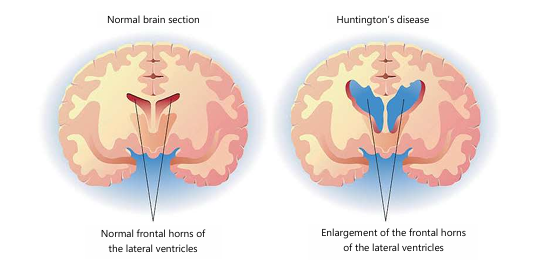
Huntington’s disease (HD) is a progressive neurodegenerative disorder with the striatum as the main location of pathology. A healthy brain's medium spiny neurons (MSNs) comprise as many as 95% projection neurons, which play a role in the feedback loop that suppresses spontaneous movements. Striatal atrophy is a hallmark of HD, and it disproportionately affects motor neuron networks (MSNs)6. Current pharmacotherapeutic approaches are based on their impact on neurotransmission in the GABA, dopamine, and glutamate systems7. Dopamine plays an essential role in controlling locomotion, perception, motivation, processing rewards, and emotions8. A large number of dopamine receptors located in both pre- and post-synaptic neurons in the brain are responsible for its action. The synaptic cleft is emptied of free dopamine by the dopamine transporter (DAT). Vesicular monoamine transporter type 2 (VMAT2) can reintroduce it into vesicles, or monoamine oxidase can degrade it into inactive metabolites7. Dopamine D1 and D2 receptors in the striatum become postsynaptically dysfunctional due to neurodegeneration in HD8 (Fig. 1). Impairment in striatal D1 and D2 receptor binding is more noticeable in manifest HD, but it is present in both premanifest and manifest HD. Executive dysfunction, diminished functional capacity, and motor impairments as determined by UHDRS-TMS have been associated in PET imaging investigations with decreased receptor binding9. An important goal of HD treatment is the regulation of dopamine levels, since it is thought that choreiform movements in HD are caused by overstimulation of dopamine receptors. Increased glutamatergic neurotransmission in thalamocortical circuits is related to hyperkinetic symptoms, and this disruption is present even in the early stages of the disease. There may be new ways to treat motor symptoms related to Huntington's disease if we focus on peripheral symptoms rather than just central neurotransmission, which include problems with the digestive, circulatory, metabolic, and muscular systems10.
|
GENETIC BASIS OF HUNTINGTON’S DISEASE
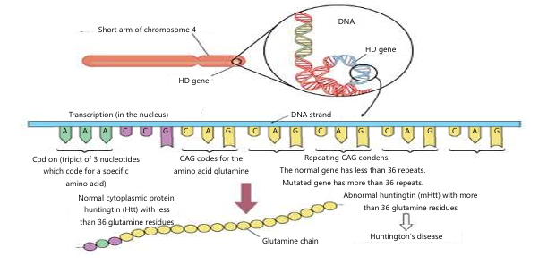
One genetic disorder caused by a single gene defect, namely a mutation in the Huntingtin (HTT) gene on chromosome 4, Huntington’s Disease is caused by a defectively expanded CAG trinucleotide repeat, making it an autosomal dominant genetic disorder, diagnosable by genetic testing11. The HTT gene synthesizes the huntingtin protein that is crucial to brain development; the exact function, however, is unknown. Mutant huntingtin, caused by the presence of the expanded CAG, affects various cellular processes, and it is this that results in Huntington’s Disease.
In unaffected people, the HTT gene has fewer than 35 repeats of the CAG triplet. 40 or more repeats of CAG are indicative of disease development, and intermediate ranges (36-39 repeats) display incomplete penetrance, with the carrier possibly displaying a milder phenotype or remaining asymptomatic for a period and displaying disease later in life12. The length of the CAG triplet can increase through the generations (this is known as genetic anticipation), especially if the mutation has been inherited from the father, because during spermatogenesis the mutation is most unstable.
Mutation and polyglutamine expansion: The defining pathological feature of HD is CAG repeat expansion within exon 1 of the HTT gene, producing a glutamine. The CAG expansion increases the length of the polyglutamine tract in the huntingtin protein, placing HD in the category of polyglutamine diseases. Instability of the elongated CAG tract is observed in both germ cells and in somatic tissues and is the reason for variability in disease presentation13. The degree of repeat expansion correlates inversely with age at onset and with age at death; bigger expansions have earlier onset and decreased survival.
Inheritance pattern: The HD is an autosomal dominant disorder. Each child of an affected individual has a 50% risk of inheriting the mutant gene, and if it is inherited, the individual is almost invariably affected because the condition has complete penetrance14. The extent of CAG repeat expansion dictates the severity and onset of HD, and a high degree of instability in the spermatogenic cycle in particular contributes to phenotypical variation with earlier onset in successive generations15,16.
PATHOPHYSIOLOGY OF HUNTINGTON’S DISEASE
HD is defined by the progressive loss of neurons in particular regions of the brain. Notably, a loss of neurons in the cerebral cortex, caudate nucleus, and putamen occurs in HD. Medium spiny neurons within the basal ganglia are particularly sensitive to degeneration and this leads to specific symptoms within the disease.
|
Chorea is thought to be caused by the loss of enkephalin-containing neurons within the indirect pathway, while dystonia and akinesia are thought to result from degeneration of substance P-containing neurons within the direct pathway. The specificity of neuronal loss in both the cortex and basal ganglia can explain the varying clinical presentation of HD patients. Several models of HD pathogenesis have been hypothesized, which occur in parallel processes. Mutant huntingtin can form intranuclear and intra-cytoplasmic inclusions that aggregate within the neurons and impede proteolytic mechanisms (Fig. 2). Aggregates of mutant huntingtin impede the ubiquitin-proteasome system, hindering protein degradation. Excitotoxicity, brought about by the hyper-release of glutamate from cortical afferents, can exacerbate neuronal damage. Mitochondrial dysfunction and energy metabolism defects, abnormal axonal transport, and changes in synaptic signaling have also all been demonstrated to occur in HD15.
PATHOGENESIS OF HUNTINGTON’S DISEASE
The pathology of HD consists of extensive neuronal loss in the striatum and cortex. The first cells affected are the medium spiny neurons, GABA- and enkephalin-containing cells that normally send signals to the lateral globus pallidus. As neuronal degeneration progresses, damage can occur throughout the basal ganglia, including the cortex and substantia nigra. The abnormal huntingtin protein causes inclusions in the cytoplasm and nucleus in the neurons. These inclusion bodies are formed by cross-linking of soluble huntingtin molecules, but it is unclear if the soluble huntingtin molecule itself is the source of toxicity or if aggregate formation is a direct cause of neuronal death. Several different neurotransmitter systems are affected in HD, but dopamine, glutamate, and GABA are the most reliably involved neurotransmitter systems and are thus considered major targets for therapeutic intervention17.
MECHANISMS AND THE ROLE OF HUNTINGTIN
The functional huntingtin is a cytoplasmic protein, carrying a polyglutamine tract within its N-terminal portion. It is ubiquitously expressed in humans and highest levels are found within the brain; it is involved in regulation of transcription, intracellular trafficking, axonal transport, neurogenesis and neuronal survival. Huntingtin expression induces the expression of neurotrophic factors (such as BDNF and NGF) vital for normal synaptic function and cognitive performance.18Lack of BDNF expression is observed in HD patients suffering from memory impairment and motor disability. Mutant mouse studies also provided the evidence that huntingtin is essential for embryonic development in the brain and its ablation abolishes neurogenesis17,18.
In HD, pathological expansion of the huntingtin polyglutamine tract occurs (in excess of 40CAG repeats forms the misfolded mutant huntingtin (mHTT))19. The misfolded protein then becomes neurotoxic in the form of accumulation of protofibrils and fibrils within the neuron. Although it may sequester some of the toxic species the structures are poorly degraded by either the ubiquitin proteasome system or the autophagy-lysosome pathway, and so remain as persistent nuclear and cytoplasmic inclusion bodies20. The striatum has been shown to have the highest levels of pathological huntingtin expression and in parallel exhibits striking selectivity for this particular part of the brain21.
The mechanisms whereby mHTT leads to neuronal dysfunction are as yet unidentified although there are several processes which contribute: Anomalous protein-protein interaction, failed proteasome and autophagy-lysosome function, transcriptional dysregulation, failure of mitochondria, oxidative stress, apoptosis, metabolic dysfunction and neuroinflammation. All contribute to a complicated cascade leading to the characteristic progressive neuronal loss seen in HD21,22.
Changes in cells: One of the most characteristic changes in HD is the formation of nuclear inclusions and cytoplasmic aggregates throughout the brain, and mHTT shows a strong propensity to form aggregates. Highly structured and largely sheet-rich amyloid fibres, highly insoluble in detergent, form polyglutamine inclusions, and a multitude of proteins, including transcription factors and protein quality control components, are recruited to polyglutamine inclusions, implying that these inclusions are detrimental23.
Macroscopic changes: The changes in the brain associated with HD are characterized by structural changes. The brain has the tendency to shrink and there is degeneration of the striatum, particularly the caudate nucleus and the putamen. The degeneration involves a loss of efferent medium spiny neurons (MSNs) and it is this loss that characterizes striatal degeneration24. The cortex also demonstrates cortical thinning but there are spatial patterns associated with the thinning of cortex and unlike the striatum, loss of cortical mass is initially seen in posterior cortical regions, progressing anteriorly over time25.
Dysregulation of the transcriptome: Transcriptional dysregulation is a major pathology and mechanism of Huntington’s disease and it results from various interconnected events. Aberrant binding of the toxic N-terminal mHTT fragment with chromatin and genomic DNA results in disruption of the transcriptional apparatus. The N-terminal polyglutamine tract in mHTT provides a motif that binds to the glutamine-rich activation domains of transcription factors CREB, Sp1, CREB-binding protein (CBP) coactivator, and dysregulates normal transcription. Striatal degeneration and cortical thinning are established structural features, and transcriptional dysregulation becomes the next level of pathology. Cortical volume loss starts from posterior to anterior of the cortex, which is in correspondence to the molecular dysregulation caused by mHTT23. Gene expression of CREB target genes is universally down-regulated in HD from experimental animal models and post-mortem studies. Among the many CREB targets, PGC-1 which plays a key role in the biogenesis of mitochondria, is down-regulated in the mouse brain as well as in post-mortem brain tissue from HD patients and connects transcriptional changes to energy deficit26.
FACTORS CONTRIBUTING TO HUNTINGTON’S DISEASE PATHOGENESIS
Oxidative stress: Oxidative stress contributes significantly to HD pathogenesis. It occurs when the body’s ability to detoxify and repair cellular damage inflicted by reactive oxygen species (ROS) becomes overwhelmed. High levels of oxidative markers have been reported in degenerating areas of the HD brain, including malondialdehyde, 8-hydroxydeoxyguanosine, 3-nitrotyrosine, and hemeoxygenase. Excessive free radical production is observed in animal models of the disease as well, adding weight to the hypothesis that oxidative damage is a key factor in the development of neuronal damage.
|
Whether oxidative damage is a causal factor or secondary effect of the degenerating cascade is unclear but the involvement of such processes is congruent with their role in many other neurodegenerative diseases2.
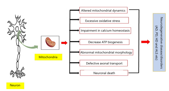
Mitochondrial dysfunction: Damage of mitochondria is also a key aspect of HD pathogenesis and occurs at the earliest stage. Studies of mitochondrial function identified mitochondrial dysfunction with defects in succinate dehydrogenase (an enzyme common to both the Krebs cycle and complex II of the ETC). Postmortem analysis of HD brain revealed a significant reduction of the complex II activity in the caudate nucleus of about 50% with respect to controls. Researchers discovered defects in the activity of complex III in the caudate nucleus and in the putamen, and in the activity of complex IV in the putamen (Fig. 3). These observations indicate that the defect is not related to only one complex but reflects a general disturbance of energy metabolism. Since most of the analyzed patients displayed severe striatal atrophy, both neuronal and glial components are likely to be responsible for these alterations27.
Excitotoxicity: Excitotoxicity contributes to neuronal death in Huntington's disease through overactivation of glutamatergic signaling, with enhanced and persistent levels of intracellular Ca2+. Stimulation of NMDA receptors by excitatory amino acids leads to opening of the mitochondrial permeability transition pore and production of free radicals, both being fatal events for the neuron. This process has been confirmed in experiments whereby administration of excitotoxins such as quinolinic acid and kainic acid directly into the striatum leads to degeneration of GABAergic medium spiny neurons, mimicking the disease process28,29.
Neuroinflammation: While inflammation in the body usually provides a protective role against harm and disease, in the case of HD, inflammation seems to play a role in driving the disease forward. Neuroinflammation is not the main cause of HD; however, there is a lot of evidence associating inflammation with disease progression. Post-mortem analyses demonstrate activated microglia and macrophages around the degenerating neurons, and increased concentrations of the pro-inflammatory cytokines IL-6, IL-1, and TNF in HD patient’s striatum and plasma as well as HD animal models. It may be that, by recognizing the mutant huntingtin aggregates as foreign material, the microglia start or increase inflammation in the brain29.
|
Apoptosis: The other process which has been implicated in HD and other long-term neurodegenerative diseases is programmed cell death, or apoptosis. Caspases, cysteine-dependent aspartate-specific proteases are important for starting and controlling the execution of the process. An up-regulation of the transcription and expression of caspase-1, caspase-3, and caspase-9 have been found in HD patients and different animal models. Furthermore, misfolded huntingtin alone can induce an apoptotic process, therefore linking protein misfolding to neuronal death30-32. New studies show the importance of gut micro flora in modulating neurologial diseases such asHD through its effects on inflammation33.
Prognosis: The diagnosis of HD is based on the characteristic symptoms and a positive family history, and has proven to exist in approximately 99% of all cases on the confirmation of the CAG trinucleotide expansion. Testing is available in three forms: Diagnostic, presymptomatic (for the investigation of a family member at risk) and prenatal, and in all circumstances extensive counselling is essential. Blood samples are always processed by DNA diagnostic laboratories so HD testing is readily available as a clinical investigation. However, it has recently been suggested that HD new mutations may be common and the usefulness of genetic testing without a family history established14.
BIOMARKERS IN HUNTINGTON’S DISEASE
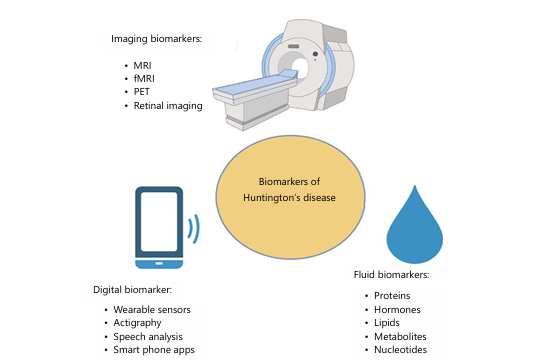
Long-term, large-scale, natural history studies such as TRACK-HD 1 and PREDICT-HD 15 have vastly enhanced our knowledge of the natural history of HD, from the premanifest state to symptom onset. In structural MRI scans, both premanifest and manifest individuals show faster rates of striatal volume loss compared to age-matched control groups with volumes significantly reduced in those more than 15 years before expected onset of disease34. Other imaging techniques such as fMRI, FDG PET, and DTI have detected abnormalities in neuronal fiber directionality and white/subcortical grey matter integrity, supporting their value as potential biomarkers (Fig. 4)35.
Behavioral and cognitive assessment techniques are also developing promising results. In the premanifest population decreased speeded tapping performance and difficulty with emotion recognition is directly related to disease progression, whereas with early HD, Stroop test performance and indirect circle tracing is correlated to disease state36. A femtomolar detection ultrasensitive single-molecule immunoassay for mHTT in CSF was established35, a marker which correlates with disease stage, as well as with the decline in motor and cognitive function. It is the biomarker under current investigation in clinical trials. Levels of tau in CSF, a recognized marker in Alzheimer's disease, have also been found to have some relevance to HD37. Recently, neurofilament light protein (NfL) has been identified as a predicting blood biomarker of HD. Elevated levels of NfL in plasma can be associated with disease onset and progress, and are easily quantifiable using a basic blood test38.
MEDICINAL PLANTS WITH PROVEN NEUROPROTECTIVE POTENTIAL
Numerous natural agents and plant-derived compounds have been tested both in vivo and in vitro, as well as in clinical settings, for their potential to prevent or treat neurologic diseases such as Huntington's disease. In many cases, their proposed mechanisms involve antioxidant and anti-inflammatory effects and may protect neurons from injury29.
A traditional Ayurvedic herb with neuroprotective and memory-improving benefits, Bacopa monnieri (BM) or Brahmi works by elevating antioxidant enzyme activities, chelation of metal ions and scavanging free radicals. It not only serves to lower stress and anxiety, but also reduces neuronal damage caused by oxidative stress. Thus, BM shows potential in HD treatment30.
Withania somnifera or Ashwagandha, has been valued in Ayurvedic medicine as an agent that promotes neuronal regeneration. It has various biological effects including anti-inflammatory, antioxidant, and neuroprotective. The active compounds, withaferin A and sitoindosides for example, increase endogenous antioxidant activity whilst diminishing lipid peroxidation. Studies indicate that Ashwagandha might work through the GABAergic system, which is linked with the pathogenesis of HD. Its root extract administered to animals showed increased cognitive function and normalized the antioxidant enzyme activity7,31,34.
One of the oldest existing trees, Ginkgo biloba has long been used in traditional medicine. Extracts from its leaves have been shown to be protective in many disease states such as psychiatric disorders, cardiovascular disease and neurodegeneration associated with aging. Antioxidant and neuroprotective effects hint at its possible utility in HD34.
Gotu kola (Centella asiatica) (CA) is an Ayurvedic Rasayana used in the enhancement of memory and prevention of aging-induced memory impairment. Experimental evidence suggests that CA can protect against toxin-induced mitochondrial dysfunction and oxidative stress by reducing the level of oxidants and restoring mitochondrial viability against toxins such as 3-nitropropionic acid, suggesting the potential use against HD-induced neurodegeneration35,36.
Ginseng, a medicinal plant, has been used for more than 2000 years in East Asia. It contains active compounds called ginsenosides and two different types (American and Asian ginseng) were tested in terms of their neuroprotective potential. Individual ginsenosides (e.g., Rb1, Rc and Rg5) have been shown to prevent excitotoxic cell death of medium spiny neurons mediated by the modulation of calcium signaling. Therefore, ginseng might act therapeutically against HD and other neurodegenerative diseases37,38.
Flavonoids belong to a class of plant polyphenols and their action in delaying neurodegeneration and offering protection against oxidative stress is well described. Flavonoids achieve neuroprotection by free radical scavenging, inhibition of nitric oxide synthase and consequently reduced peroxynitrite-mediated damage. Similar to the neuroprotection demonstrated in both Parkinson’s and Alzheimer’s diseases, flavonoids may help the HD brain to resist oxidative damage and neurological decline39,40.
CURRENT CLINICAL STRATEGIES
Existing therapies are aimed at the symptoms and do not reverse or halt disease progression. Tetrabenazine (TBZ; Xenazine) was approved in 2008 by the FDA for use as a treatment for chorea in Huntington's patients. Most recently, the FDA has approved deutetrabenazine (AUSTEDO), a deuterated form of TBZ, for the treatment of tardive dyskinesia and chorea in Huntington's disease patients. The deuterated form of the drug possesses a more favourable pharmacokinetic profile, thus improving its tolerance and effectiveness40.
CHOREA MANAGEMENT
TBZ is a Vesicular Monoamine Transporter type 2 (VMAT2) inhibitor that causes decreased release of dopamine into the synaptic terminal, thereby decreasing binding of dopamine to postsynaptic dopamine receptors. Diminution of dopaminergic signaling leads to antichoreic effects and control of involuntary movement disorders in HD. Studies in animal models have demonstrated that TBZ treatment diminishes chorea, striatal neuronal loss, and motor deficits. Thus, TBZ is an FDA-approved drug for chorea41.
ADVANCES WITH DEUTETRABENAZINE
One of the shortcomings of TBZ therapy, despite its efficacy, is that it has a short half-life and is metabolized unevenly, resulting in the need for administration three times per day with difficulty in reaching steady state. These pharmacokinetic shortcomings lead to adverse effects such as akathisia, nausea, and anxiety. These problems were overcome in deutetrabenazine, which is identical to TBZ, but the hydrogens were replaced by deuterium atoms, strengthening the bonds and retarding metabolism, prolonging the half-life so that it can be administered at less frequent doses, usually twice daily. In clinical trials, deutetrabenazine was found to result in an equivalent improvement in chorea compared with TBZ, and further proved advantageous in the assessment of dystonia and total motor score. Crucially, unlike TBZ therapy, which can result in adverse symptoms as a consequence of dopamine deficiency, it did not produce symptoms associated with Parkinsonism or depression which are important considerations with such drugs in HD where depression and suicide are so prevalent. It was approved by the FDA in 2017 for treatment of chorea in HD36,37.
CHALLENGES IN CURRENT CLINICAL MANAGEMENT
While therapies targeting symptoms of chorea, such as tetrabenazine and deutetrabenazine, have improved clinical management, they do not impact the overall course of Huntington’s Disease. Individuals with HD still encounter psychiatric disorders, cognitive decline and motor deficits which lead to loss of function and quality of life. Disease onset, the degree of symptoms and progression vary across individuals which makes the measurement of therapeutic effects in clinical trials challenging when testing heterogeneous populations of patients. A major impediment to treating and curing HD is the lack of accurate biomarkers to both identify disease onset early, as well as to provide an objective measure to track progression in HD which limits intervention in affected individuals.
Pharmacological treatments can be accompanied by other risks. While effective for treating chorea, VMAT2 inhibitors have known psychiatric adverse effects which can include depression, anxiety and akathisia. This is a concern considering the significantly increased rate of suicide associated with HD. In addition to chorea, psychiatric features such as irritability, apathy and psychosis commonly occur in individuals with HD. The management of these symptoms is complex, and the currently available pharmacotherapy options do not adequately treat these conditions without severe side effects34.
FUTURE CLINICAL INSIGHTS
CRISPR-Cas9 gene editing: The most exciting potential benefit of CRISPR-Cas9 is its ability to target directly expanded CAG repeats on the HTT gene. In preclinical trials, removing or silencing the mutant huntingtin has resulted in reduced toxic aggregation of protein and enhanced neuronal viability. If possible, this method has the potential to stop or even reverse the disease process. Potential hurdles include the possibility of off-target gene alterations, difficulty crossing the blood-brain barrier with CRISPR components, and moral implications regarding the editability of germline cells. Current research is aiming to improve delivery methods (using viral vector or nanoparticle) for safe and accurate delivery37,38.
RNA-based therapies: RNA-based therapies such as antisense oligonucleotides (ASOs) and RNA interference (RNAi) seek to lower levels of mutant huntingtin protein. Clinical studies of ASOs such as tominersen have reported suppression of huntingtin expression but questions regarding long-term safety, immunogenicity and optimal dosing have inhibited translation to the clinic. Future steps forward include the design of more specific, less toxic next-generation ASOs and combination therapy approaches using RNA-based drugs and neuroprotective therapies39,40.
Stem cell and neuroregenerative approaches: Stem cell transplantation and neurotrophic factor delivery are being investigated as strategies to restore neuronal populations and promote brain repair. Early studies suggest improvements in motor function and neuronal survival, but challenges such as immune rejection, integration into existing neural circuits, and scalability remain unresolved. Advances in induced pluripotent stem cells (iPSCs) and gene-edited stem cell lines may help overcome these barriers, offering personalised regenerative therapies tailored to individual patients41,42.
Biomarkers and precision medicine: The discovery of accurate biomarkers is vital for progress toward future HD treatment strategies. Neuroimaging, specifically MRI and diffusion tensor imaging, may identify the earliest changes in structure and function before the onset of clinical signs. Analysis of cerebrospinal fluid to detect mutant huntingtin and tau proteins, and plasma levels of neurofilament light protein (NfL) are being refined as objective biomarkers for disease inception and advancement and are important for future use in "personalized medicine" applications, allowing the personalization of treatments43.
CONCLUSION
Huntington’s disease (HD) is an autosomal dominantly inherited neurodegenerative disorder that predominantly affects striatal medium spiny neurons and is characterized by progressive motor impairments, cognitive deterioration and psychiatric disturbances. HD is caused by an expanded polyglutamine (polyQ) tract within the HTT gene that produces toxic mutant huntingtin (mHTT) protein. Genetic testing is widely available, but it may only give a positive result and the psychological and social implications for both the patient and their family mean that genetic counseling must occur beforehand. Treatment is currently aimed at the management of symptoms (chorea, mental disorders and Parkinsonism) not disease modifying. The discovery of VMAT 2 inhibitors such as tetrabenazine and deutetrabenazine have led to improved motor symptom control but the presence of psychiatric side effects and the non-disease modifying nature are significant drawbacks. Studies have repeatedly shown that oxidative stress plays a pivotal role in the pathogenesis of HD causing the neurons to be susceptible to degeneration. In addition, studies have also shown that there are both in vitro and in vivo benefits of antioxidants and neuroprotective substances from herbal plants (Bacopa monnieri, Withania somnifera, ginseng) which could perhaps be of benefit as adjunct therapy in managing symptoms. Looking ahead to future clinical implications, gene targeted therapies including CRISPR-Cas9, RNA directed technologies, and stem cell therapies offer hopeful solutions targeting the genetic underpinnings of HD, and may change treatments beyond symptom management toward a disease modifying approach. Along with genetic therapies, successful biomarkers like neurofilament light protein (NfL) and innovative imaging technologies will play an integral role in early diagnosis and tracking progress to individualize treatment plans. While a cure for HD does not exist currently, future clinical approaches combining gene targeted therapies with neuroprotectants and biomarkers will enable precision medicine and can significantly impact clinical care toward an emphasis on altering disease course and impacting the lives of individuals with HD and their families.
SIGNIFICANCE STATEMENT
Huntington’s disease is a devastating inherited neurodegenerative disorder for which effective disease-modifying treatments remain unavailable. This review integrates current evidence on the molecular and biochemical mechanisms underlying neuronal dysfunction, including excitotoxicity, mitochondrial impairment, oxidative stress, and altered cellular signaling. By highlighting emerging therapeutic strategies such as RNA-targeted therapies, CRISPR-Cas9 gene editing, stem cell-based approaches, and antioxidant interventions, this review identifies promising directions for future research. The findings may support improved understanding of Huntington’s disease pathogenesis, facilitate the development of targeted therapies, and encourage greater attention to diagnosis, surveillance, and equitable access to care, particularly in underrepresented populations.
ACKNOWLEDGMENT
We thank all the researchers who contributed to the success of this research project.
REFERENCES
- Tabrizi, S.J., D.R. Langbehn, B.R. Leavitt, R.A.C. Roos and A. Durr et al., 2009. Biological and clinical manifestations of Huntington's disease in the longitudinal TRACK-HD study: Cross-sectional analysis of baseline data. Lancet Neurol., 8: 791-801.
- Zheng, Z. and M.I. Diamond, 2012. Huntington Disease and the Huntingtin Protein. In: Progress in Molecular Biology and Translational Science, Teplow, D.B. (Ed.), Elsevier, Amsterdam, Netherlands, ISBN: 978-0-12-385883-2, pp: 189-214.
- Yohrling, G., K. Raimundo, V. Crowell, D. Lovecky, L. Vetter and L. Seeberger, 2020. Prevalence of Huntington's disease in the US (954). Neurology, 94.
- Coppen, E.M. and R.A.C. Roos, 2017. Current pharmacological approaches to reduce chorea in Huntington's disease. Drugs, 77: 29-46.
- Jankovic, J. and R.A.C. Roos, 2014. Chorea associated with Huntington's disease: To treat or not to treat? Movement Disord., 29: 1414-1418.
- Ribaï, P., K. Nguyen, V. Hahn-Barma, I. Gourfinkel-An and M. Vidailhet et al., 2007. Psychiatric and cognitive difficulties as indicators of juvenile Huntington disease onset in 29 patients. Arch. Neurol., 64: 813-819.
- Frank, S., 2014. Treatment of Huntington's disease. Neurotherapeutics, 11: 153-160.
- André, V.M., C. Cepeda and M.S. Levine, 2010. Dopamine and glutamate in Huntington's disease: A balancing act. CNS Neurosci. Ther., 16: 163-178.
- Andrews, T.C., R.A. Weeks, N. Turjanski, R.N. Gunn and L.H.A. Watkins, 1999. Huntington's disease progression: PET and clinical observations. Brain, 122: 2353-2363.
- Carroll, J.B., G.P. Bates, J. Steffan, C. Saft and S.J. Tabrizi, 2015. Treating the whole body in Huntington's disease. Lancet Neurol., 14: 1135-1142.
- Cattaneo, E., C. Zuccato and M. Tartari, 2005. Normal huntingtin function: An alternative approach to Huntington's disease. Nat. Rev. Neurosci., 6: 919-930.
- Langbehn, D.R., R.R. Brinkman, D. Falush, J.S. Paulsen and M.R. Hayden, 2004. A new model for prediction of the age of onset and penetrance for Huntington's disease based on CAG length. Clin. Genet., 65: 267-277.
- Wheeler, V.C., F. Persichetti, S.M. McNeil, J.S. Mysore and S.S. Mysore et al., 2007. Factors associated with HD CAG repeat instability in Huntington disease. J. Med. Genet., 44: 695-701.
- Myers, R.H., 2004. Huntington's disease genetics. Neurotherapeutics, 1: 255-262.
- Bates, G., 2003. Huntingtin aggregation and toxicity in Huntington's disease. Lancet, 361: 1642-1644.
- Ajitkumar, A., F. Lui and O. de Jesus 2025. Huntington Disease. StatPearls Publishing, Treasure Island, Florida.
- Arrasate, M., S. Mitra, E.S. Schweitzer, M.R. Segal and S. Finkbeiner, 2004. Inclusion body formation reduces levels of mutant huntingtin and the risk of neuronal death. Nature, 431: 805-810.
- Zuccato, C. and E. Cattaneo, 2009. Brain-derived neurotrophic factor in neurodegenerative diseases. Nat. Rev. Neurol., 5: 311-322.
- Zeitlin, S., J.P. Liu, D.L. Chapman, V.E. Papaioannou and A. Efstratiadis, 1995. Increased apoptosis and early embryonic lethality in mice nullizygous for the Huntington's disease gene homologue. Nat. Genet., 11: 155-163.
- Borrell-Pagès, M., D. Zala, S. Humbert and F. Saudou, 2006. Huntington's disease: From huntingtin function and dysfunction to therapeutic strategies. Cell. Mol. Life Sci., 63: 2642-2660.
- Gil, J.M. and A.C. Rego, 2008. Mechanisms of neurodegeneration in Huntington's disease. Eur. J. Neurosci., 27: 2803-2820.
- Saudou, F., S. Finkbeiner, D. Devys and M.E. Greenberg, 2004. Huntingtin acts in the nucleus to induce apoptosis but death does not correlate with the formation of intranuclear inclusions. Cell, 95: 55-66.
- Labbadia, J. and R.I. Morimoto, 2013. Huntington's disease: Underlying molecular mechanisms and emerging concepts. Trends Biochem. Sci., 38: 378-385.
- Jamwal, S. and P. Kumar, 2015. Antidepressants for neuroprotection in Huntington's disease: A review. Eur. J. Pharmacol., 769: 33-42.
- Chen, C.M., 2011. Mitochondrial dysfunction, metabolic deficits, and increased oxidative stress in Huntington's disease. Chang Gung Med. J., 34: 135-152.
- Kumar, A., M. Vaish and R.R. Ratan, 2014. Transcriptional dysregulation in Huntington's disease: A failure of adaptive transcriptional homeostasis. Drug Discovery Today, 19: 956-962.
- Quintanilla, R.A. and G.V.W. Johnson, 2009. Role of mitochondrial dysfunction in the pathogenesis of Huntington's disease. Brain Res. Bull., 80: 242-247.
- diFiglia, M., 1990. Excitotoxic injury of the neostriatum: A model for Huntingtons disease. Trends Neurosci., 13: 286-289.
- Kalonia, H., P. Kumar and A. Kumar, 2011. Licofelone attenuates quinolinic acid induced Huntington like symptoms: Possible behavioral, biochemical and cellular alterations. Prog. Neuro-Psychopharmacol. Biol. Psychiatry, 35: 607-615.
- Friedlander, R.M., 2003. Apoptosis and caspases in neurodegenerative diseases. N. Engl. J. Med., 348: 1365-1375.
- Leegwater-Kim, J. and J.H.J. Cha, 2004. The paradigm of Huntington’s disease: Therapeutic opportunities in neurodegeneration. Neurotherapeutics, 1: 128-138.
- Li, S.H., S. Lam, A.L. Cheng and X.J. Li, 2000. Intranuclear huntingtin increases the expression of caspase-1 and induces apoptosis. Hum. Mol. Genet., 9: 2859-2867.
- Kanhere, H.S., Y.U. Rahangdale, A.S. Bodele, D.I. Wadhwani, A.R. Ghoshewar and S.P. Karande, 2021. Neurological disorders associated with impaired gut microbiota. GSC Biol. Pharm. Sci., 15: 029-039.
- Aylward, E.H., P.C. Nopoulos, C.A. Ross, D.R. Langbehn and R.K. Pierson et al., 2011. Longitudinal change in regional brain volumes in prodromal Huntington disease. J. Neurol. Neurosurg. Psychiatry, 82: 405-410.
- Harrington, D.L., M.M. Smith, Y. Zhang, N.E. Carlozzi and J.S. Paulsen, 2012. Cognitive domains that predict time to diagnosis in prodromal Huntington disease. J. Neurol. Neurosurg. Psychiatry, 83: 612-619.
- Ross, C.A., E.H. Aylward, E.J. Wild, D.R. Langbehn and J.D. Long et al., 2014. Huntington disease: Natural history, biomarkers and prospects for therapeutics. Nat. Rev. Neurol., 10: 204-216.
- Mfon, N.P., B.E. Obi, M.C. Chuwang, F. Nkemehule and M.A. Oladosu et al., 2025. Evaluation of the effects of Cannabis sativa on the body weight and uterus of female albino rats. Trends Med. Res., 20: 44-49.
- Eze, M.N., H.E. Odoh and M.C. Apeh, 2023. Assessment of phytochemicals and antioxidant potential of the stem bark of Albizia chevalieri. Bioscientist J., 11: 43-56.
- Abah, M.A., M.A. Oladosu, O.K. Elijah, E.P. Chikelum and A.B. Olanrewaju et al., 2025. The evolving role of sodium-glucose linked transporter-2 (SGLT2) inhibitors in heart failure management: A paradigm shift. Open J. Cardiol. Heart Dis., 5.
- Okwah, M.N., M.A. Abah, M.A. Oladosu, C.T. Blessing, A.I. Anku and O.D. Agida, 2026. Anti-inflammatory effects of omega-3 fatty acids: Evidence from circulating biomarkers and inflammatory gene-expression studies. Afr. Res. Rep., 2: 365-382.
- Oluwatobi, O.F., A. Fatai, O.M. Abimbola, M.A. Abah and I.E. Peter et al., 2026. Computational screening of bioactive compounds targeting dihydrofolate reductase in Trypanosoma brucei for inhibitory activity. Res. J. Parasitol., 21: 9-12.
- Abah, M.A., N.A. Cynthia, M.A. Oladosu, N.R. Yohanna, E.T. Ejim and O. Oghenekenu, 2026. Advances in liquid biopsy for early cancer detection: A comprehensive review of circulating tumor DNA and other biomarkers. Acta Pharma Rep., 5: 19-27.
- Oladosu, M.A., M.A. Abah, S.O. Julius, O.A. Balogun and A. Olalekan, 2026. The role of autophagy in cell survival, aging, and disease. J. Cancer Res. Rev., 3: 66-80.
How to Cite this paper?
APA-7 Style
Abah,
M.A., Abimbola,
M.O., Yohanna,
N.R., Ola,
A.A., Akinlabi,
A.O., Rahima,
Y. (2026). Molecular Mechanisms and Biochemical Alterations in Huntington’s Disease: Current Perspectives and Emerging Insights. Trends in Medical Research, 21(1), 64-76. https://doi.org/10.3923/tmr.2026.64.76
ACS Style
Abah,
M.A.; Abimbola,
M.O.; Yohanna,
N.R.; Ola,
A.A.; Akinlabi,
A.O.; Rahima,
Y. Molecular Mechanisms and Biochemical Alterations in Huntington’s Disease: Current Perspectives and Emerging Insights. Trends Med. Res 2026, 21, 64-76. https://doi.org/10.3923/tmr.2026.64.76
AMA Style
Abah
MA, Abimbola
MO, Yohanna
NR, Ola
AA, Akinlabi
AO, Rahima
Y. Molecular Mechanisms and Biochemical Alterations in Huntington’s Disease: Current Perspectives and Emerging Insights. Trends in Medical Research. 2026; 21(1): 64-76. https://doi.org/10.3923/tmr.2026.64.76
Chicago/Turabian Style
Abah, Moses, Adondua, Micheal Oladosu Abimbola, Nathan Rimamsanati Yohanna, Abdulkabir Abdulquadri Ola, Ahmed Olajide Akinlabi, and Yunusa Rahima.
2026. "Molecular Mechanisms and Biochemical Alterations in Huntington’s Disease: Current Perspectives and Emerging Insights" Trends in Medical Research 21, no. 1: 64-76. https://doi.org/10.3923/tmr.2026.64.76

This work is licensed under a Creative Commons Attribution 4.0 International License.
