
Effect of Fixative Type and Duration on Staining Quality in Wistar Rat Testicular Tissue
-
Babatunde Oluwaseun Ibitoye


Department of Human Anatomy, Federal University of Technology Akure, Ondo State, Nigeria
Adesua Emmanuel OgunmokunwaDepartment of Human Anatomy, Faculty of Basic Medical Sciences, Achievers University, Owo, Ondo State, Nigeria
Tunde Temidayo ElemosoDepartment of Human Anatomy, Faculty of Basic Medical Sciences, Achievers University, Owo, Ondo State, Nigeria
Francis Temitope AdeniranDepartment of Human Anatomy, Faculty of Basic Medical Sciences, Achievers University, Owo, Ondo State, Nigeria
Philip Ibukunoluwa AjayiDepartment of Human Anatomy, Federal University of Technology Akure, Ondo State, Nigeria
| Received 17 Dec, 2025 |
Accepted 02 Jun, 2026 |
Published 23 Jun, 2026 |
Background and Objective: Fixatives and fixation protocols are critical determinants of histological staining quality, especially for sensitive tissues like the testis. However, comprehensive comparative evaluations of commonly used fixatives and fixation durations remain limited. This study aimed to assess the effects of four fixatives-Bouin’s fluid, Davidson’s fluid, formalin, and neutral buffered formalin (NBF)-at three fixation durations (48 hours, 7 days, and 4 weeks) on the staining fidelity of Wistar rat testicular tissue using different histological techniques. Materials and Methods: Testicular tissues from Wistar rats were fixed using the specified fixatives for each duration. Sections were stained with Hematoxylin and Eosin (H&E), Gordon and Sweet (G&S), and Periodic Acid-Schiff (PAS) techniques. Staining quality was evaluated based on fixation effectiveness, tissue integrity, and artefact presence. Data were analyzed using descriptive statistics and appropriate comparative tests at p<0.05. Results: Bouin’s fluid yielded superior results for H&E and G&S at 48 hours but performed poorly with PAS staining. Davidson’s fluid maintained consistent G&S staining across all durations and demonstrated high initial PAS quality. Formalin showed optimal H&E staining at 48 hours but declined with prolonged fixation. The NBF was moderately effective for short-term H&E but lacked versatility across staining methods. Conclusion: This study emphasizes the importance of selecting appropriate fixatives and fixation durations tailored to specific staining techniques. Bouin’s and Davidson’s fluids are preferable for short- and long-term use respectively, depending on the staining method. These findings can guide histologists and researchers in optimizing testicular tissue processing for enhanced diagnostic and research accuracy.
| Copyright © 2026 Ibitoye et al. This is an open-access article distributed under the Creative Commons Attribution License, which permits unrestricted use, distribution, and reproduction in any medium, provided the original work is properly cited. |
INTRODUCTION
Fixatives and fixation protocols are critical in histological and histopathological studies, as they ensure the structural and molecular integrity of tissues for subsequent staining and microscopic evaluation1. The choice of fixative and the duration of fixation significantly influence the quality and diagnostic value of stained tissue sections2. This is particularly relevant for specialized staining techniques such as Hematoxylin and Eosin (H&E), Gordon and Sweet (G&S), and Periodic Acid-Schiff (PAS), which are widely used for studying morphological, connective tissue, and carbohydrate-related structures, respectively.
Testicular tissue presents unique challenges for histological assessment due to its complex architecture and sensitivity to handling and fixation conditions3. Fixatives such as Bouin’s fluid, Davidson’s fluid, formalin, and neutral buffered formalin (NBF) are commonly employed for preserving testicular tissues4. However, the efficacy of these fixatives varies based on their chemical composition and their ability to stabilize specific cellular and extracellular components4. For instance, Bouin’s fluid, known for its picric acid and acetic acid components, provides excellent nuclear and cytoplasmic contrast but may alter glycogen-rich structures5. In contrast, formalin-based fixatives are widely used due to their availability and cost-effectiveness but are less effective for preserving delicate structures over extended durations6.
The fixation time further complicates the choice of fixative, as prolonged fixation can result in over-hardening, shrinkage, and loss of antigenicity, while insufficient fixation leads to poor staining outcomes and artifacts7. These factors are especially critical for techniques like G&S, which rely on the preservation of argentophilic structures, and PAS, which depends on intact carbohydrate moieties8. Despite the critical role of fixation in tissue preparation, limited studies comprehensively compare the combined effects of fixative types and fixation durations on multiple staining techniques for testicular tissues9.
This study aims to evaluate the effects of four commonly used fixatives (Bouin’s fluid, Davidson’s fluid, formalin, and NBF) and varying fixation durations (48 hours, 7 days, and 4 weeks) on the quality of H&E, G&S, and PAS staining in testicular tissue of Wistar rats. The findings will provide insights into optimizing fixation protocols for testicular histology, with implications for both research and diagnostic practices.
MATERIALS AND METHODS
Study area: This study was conducted between February and March 2024 at the Animal House of the Department of Anatomy, Federal University of Technology, Akure, Nigeria.
Experimental design: This study was designed to assess the effects of four fixatives (Bouin’s fluid, Davidson’s fluid, formalin, and neutral buffered formalin and three fixation durations (48 hours, 7 days, and 4 weeks) on the staining quality of testicular tissues in adult male Wistar rats using Hematoxylin and Eosin (H&E), Gordon and Sweet (G&S), and Periodic Acid-Schiff (PAS) staining techniques.
Animal model: A total of 15 adult male Wistar rats, weighing between 200 to 300 g were used for this study. The animals were housed in standard laboratory conditions with a 12-hour light/dark cycle, adequate ventilation, and access to food and water ad libitum for 2 weeks.
Tissue collection: The Wistar rats were anaesthetised with 50 mg/kg of ketamine, and the testes were excised by making a midline incision in the abdomen, deepened to the peritoneum in the pelvic region, to expose the testes, which were then immediately rinsed in cold phosphate-buffered saline (PBS) to remove blood and debris10.
Fixation protocol: The testicular tissues were randomly divided into four groups based on the fixative used:
| • | Bouin’s fluid | |
| • | Davidson’s fluid | |
| • | Formalin (10%) | |
| • | Neutral Buffered Formalin (10%) |
| Table 1: | Semi-quantitative evaluation of staining quality of testicular tissue using self-designed scale | |||
| Criteria/score | 0-2 | 3-4 | 5-6 | 7-8 | 9-10 | ||
| Cellular details | Poor visibility of cellular details | Vague cellular outlines | Fair resolution of structural details | Good visibility of key structures | Crisp cellular morphology; robust | ||
| Score | 0 | 1 | 2 | 3 | 4 | 5 | |
| Fixation quality |
No fixation; tissue/cells are degraded or autolyzed |
widespread shrinkage, poor preservation of morphology |
Suboptimal fixation; partial preservation, some cytoplasmic and nuclear distortion |
Moderate fixation; structures generally visible but with some artifacts |
Good fixation; well- preserved morphology with minor flaws |
Excellent fixation; uniform, artifact-free preservation of cells/tissues |
|
| Staining quality |
No staining or extremely poor contrast |
Incomplete or uneven staining; difficult to interpret |
Weak staining; some structures are difficult to differentiate |
Adequate staining; most structures are visible with effort |
Good staining; clear contrast, minor inconsistencies |
Optimal staining; high contrast, all features well- demarcated |
|
| Score | 0 | 1 | 2 | 3 | |||
| Artifact presence |
Numerous or large artifacts impairing interpretation |
Moderate artifacts present, but interpretation still possible |
Few minor artifacts; interpretation largely unaffected |
No observable artifacts; slide is clean and interpretable |
|||
Each group was further subdivided into three subgroups corresponding to fixation durations of 48 hours, 7 days, and 4 weeks. Tissues were immersed in their respective fixatives in labeled containers and stored at room temperature under appropriate conditions.
Tissue processing: After fixation, tissues were dehydrated through graded ethanol series, cleared in xylene, and embedded in paraffin wax. Paraffin blocks were sectioned at 5 μm thickness using a rotary microtome11.
Staining techniques: The following staining techniques were performed on tissue sections:
| • | Hematoxylin and Eosin (H&E): For general tissue morphology and nucleocytoplasmic contrast12 | |
| • | Gordon and Sweet (G&S): For the demonstration of reticular and connective tissue fibers | |
| • | Periodic Acid-Schiff (PAS): For carbohydrate-rich structures and basement membranes13 |
All staining procedures were conducted according to standard protocols, with strict adherence to reagent preparation and timing.
Semi-quantitative evaluation of staining quality: The stained slides were evaluated under a light microscope by three independent observers who are experienced anatomists, blinded to the experimental groups. Staining quality was scored on a self-designed scale with a minimum score of 0 and a maximum of 23. The scoring criteria and values are in Table 1.
Statistical analysis: Data were analyzed using GraphPad Prism version 8.0 and expressed as Mean±Standard Deviation. Comparisons were made using Two-way ANOVA. A p<0.05 was considered statistically significant.
Ethical considerations: All experimental procedures adhered to the guidelines of the International Animal Care and Use Committee (IACUC), ensuring humane treatment of animals and minimizing distress during handling and euthanasia. Ethical approval was obtained from the Ethical Committee of the Federal University of Technology, Akure, under the approval number FUTA/ETH/24/23.
RESULTS
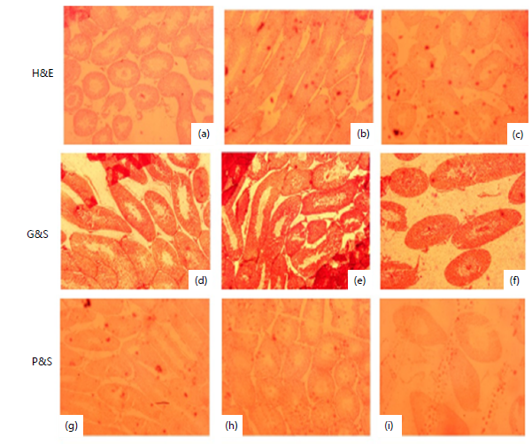
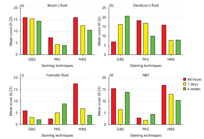
Bouin’s fluid: Bouin’s fluid preserved testicular morphology effectively at 48 hours, with high staining scores in H&E and G&S. However, PAS staining was consistently poor across all time points. Staining quality declined progressively at 7 days and 4 weeks, indicating Bouin’s fluid is best suited for short-term fixation, particularly for H&E and G&S (Fig. 1(a-i), 5a). This was supported by a significant time-dependent effect (F = 7.98, p = 0.0402).
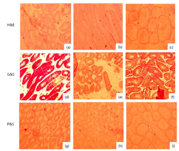
Davidson’s fluid: Davidson’s fluid produced optimal H&E and PAS staining at 48 hours, with reduced quality at 7 days and 4 weeks. In contrast, G&S staining improved progressively, peaking at 4 weeks, indicating enhanced preservation of connective tissue elements over time. These trends, illustrated in (Fig. 2(a-i), 5b), showed no statistically significant differences across durations (F = 0.02, p = 0.9851), suggesting time-stable but stain-specific effects.
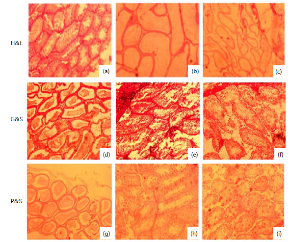
Formalin: Formalin fixation resulted in high H&E staining quality at 48 hours, which declined sharply at 7 days and 4 weeks, indicating reduced morphological preservation over time. G&S staining was consistently poor, while PAS staining showed slight improvement with prolonged fixation. As shown in (Fig. 3(a-i), 5c), these patterns were not statistically significant (F = 7.98, p = 0.6308), suggesting limited long-term efficacy across staining techniques.
|
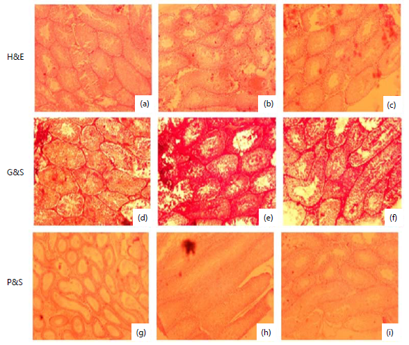
Neutral buffered formalin: The NBF showed strong initial H&E staining at 48 hours, with gradual decline at 7 days and 4 weeks. A G&S staining was moderate at 48 hours, dropped at 7 days, and slightly improved at 4 weeks. PAS staining remained poor across all durations. As illustrated in (Fig. 4(a-i), 5d), these changes were not statistically significant (F = 1.70, p = 0.2918), indicating limited consistency over time.
DISCUSSION
Bouin’s fluid showed distinct patterns in its effects on different staining techniques (H&E, G&S, and PAS) based on fixation time. The staining quality with Bouin's fluid was initially high (16 score at 48 hours), indicating its excellent ability to preserve tissue morphology and maintain nucleocytoplasmic contrast. Over time, the staining quality decreased slightly (15.5 score at 7 days and 14.5 score at 4 weeks), suggesting a gradual decline in fixation effectiveness for prolonged durations.
|
This trend aligns with studies highlighting Bouin’s as ideal for short-term preservation due to its picric acid content, which provides sharp nuclear staining14 (Fig. 1(a-i), 5a). The performance of Bouin's fluid for G&S staining showed a similar pattern. At 48 hours, the staining quality was moderate (16 score), but a decline was observed at 7 days (15.5 score) and 4 weeks (14.5 score). This indicates that while Bouin’s provides good initial preservation, the fixative may not support the prolonged structural integrity required for silver-based staining. Kiernan et al.15 corroborated this, noting that Bouin’s fixation could result in loss of fine structural details over time due to protein denaturation (Fig. 1(a-i), 5a).
|
Bouin’s showed the poorest performance for PAS staining (7.5 at 48 hours, 4.5 at 7 days, and 4 at 4 weeks). This highlights its inadequacy in preserving polysaccharides and other PAS-reactive substances. The picric acid component may interfere with carbohydrate-rich structures, reducing staining intensity. This finding aligns with Buesa and Peshkov16, who found Bouin’s less effective for glycogen-rich tissues, especially after extended fixation durations (Fig. 1(a-i), 5a). Davidson’s fluid demonstrated robust preservation and staining outcomes across techniques and durations. The H&E staining was initially high at 48 hours (16 score), but it decreased significantly at 7 days (8) and 4 weeks (8). Davidson’s fluid contains acetic acid, which enhances nuclear detail, but prolonged fixation may lead to over-hardening, reducing staining intensity.
|
This is consistent with Mills17, who noted Davidson’s excellent performance for short-term H&E staining but suggested time-sensitive optimization (Fig. 2(a-i), 5b). G&S staining showed a unique improvement over time (7 at 48 hours, 16.5 at 7 days, and 21 at 4 weeks). This suggests that Davidson’s fluid stabilizes components relevant to silver staining during prolonged fixation. Carson et al.18 observed a similar trend, attributing the improvement to reduced tissue artifacts and enhanced impregnation efficiency over time (Fig. 2(a-i), 5b). Davidson’s provided the highest PAS staining intensity at 48 hours (18.5), but a decline was observed at 7 days (17) and 4 weeks (10). While its initial performance is attributed to effective carbohydrate preservation, the gradual decline over time might result from fixative-tissue interactions that degrade PAS-reactive components (Fig. 2(a-i), 5b).
|
Formalin showed variable effects depending on the staining technique and duration. A H&E staining was most effective at 48 hours (17.5), but it decreased significantly at 7 days (7) and 4 weeks (4). This aligns with its known limitations, where prolonged fixation causes over-hardening and reduced staining quality due to extensive crosslinking. A Fox et al.19 reported similar findings, emphasizing formalin’s suitability for short-term fixation in routine H&E staining (Fig. 3(a-i), 5c). The G&S staining performance was poor across all durations (6 at 48 hours, 3 at 7 days, and 2 at 4 weeks). Formalin's inability to preserve structures necessary for silver staining likely accounts for this decline. This trend is supported by Kiernan15, who noted that formalin's reactive aldehyde groups interfere with argentophilic components (Fig. 3(a-i), 5c). PAS staining performance was initially poor (2.5 at 48 hours) but improved slightly over time (5 at 7 days and 9 at 4 weeks). This unusual trend may be due to secondary crosslinking effects stabilizing carbohydrate-rich structures over prolonged fixation. Buesa and Peshkov16 noted that while formalin is not the best choice for PAS, extended fixation could improve staining outcomes with optimization (Fig. 3(a-i), 5c). The NBF provided moderate results across staining techniques but varied significantly with duration. The NBF showed strong initial staining (17 at 48 hours), with a decline over time (13 at 7 days, 10.5 at 4 weeks). This suggests good nucleocytoplasmic preservation initially but reduced quality with prolonged fixation, similar to plain formalin. Mills17 observed comparable trends, recommending NBF for short-term fixation in H&E protocols (Fig. 4(a-i), 5d). A G&S staining was moderate at 48 hours (15.5) but declined sharply at 7 days (6.5) before slightly improving at 4 weeks (14). The decline at 7 days suggests tissue artifacts, while the improvement at 4 weeks may be due to secondary stabilization of silver-reactive structures (Fig. 4(a-i), 5d). The PAS staining was poor across all durations (3 at 48 hours, 2 at 7 days, and 4.5 at 4 weeks). This indicates that NBF, like formalin, is unsuitable for preserving PAS-reactive substances, particularly for long-term fixation (Fig. 4(a-i), 5d).
CONCLUSION
Each fixative displayed unique interactions with staining techniques and fixation times. Bouin’s and Davidson’s fluids excelled for short-term preservation, particularly for H&E and PAS, while Davidson’s proved superior for G&S over extended periods. Formalin and NBF, although widely used, demonstrated limited versatility and suboptimal results for specialized staining protocols like PAS and G&S. The findings emphasize the importance of tailoring fixative and fixation duration to specific histological objectives.
SIGNIFICANCE STATEMENT
This study provides comparative evidence on the influence of fixative type and fixation duration on the staining quality of Wistar rat testicular tissue using H&E, Gordon and Sweet, and PAS techniques. The findings highlight that appropriate selection of fixatives and fixation periods significantly affects tissue preservation, staining fidelity, and histological interpretation. The study offers practical guidance for histologists, anatomists, and researchers in optimizing fixation protocols for improved diagnostic accuracy and experimental reliability in testicular tissue studies.
ACKNOWLEDGMENT
The authors sincerely thank the Department of Human Anatomy, Federal University of Technology, Akure, and Achievers University, Owo, for providing the laboratory facilities and technical support necessary for the successful completion of this research.
REFERENCES
- Dağdeviren, T., H.K. Yolcu and B. Ünver, 2024. Histological fixation process and fixatives. Turk. J. Agric. Food Sci. Technol., 12: 1482-1486.
- Bamisi, O.D. and M.O. Alese. 2020. Effects of various fixatives and temperature on the quality of glycogen demonstration in the brain and liver tissues. Ann. Diagn. Pathol., 48.
- Tian, P., Z. Yang, C. Qu, X. Qi, L. Zhu, G. Hao and Y. Zhang, 2024. Exploration of tissue fixation methods suitable for digital pathological studies of the testis. Eur. J. Med. Res., 29.
- Feng, X.Z., W.Y. Wang, T. Gong and L.J. Meng, 2021. Different fixatives influence morphology, antigen preservation, and TUNEL staining in chicken (Gallus gallus) testis. Acta Histochem., 123.
- Adel Kareem, D. and M.F. Majeed, 2024. A brief overview of the role and function of fixative and fixation: Review. Univ. Thi-Qar J. Agric. Res., 13: 264-271.
- Ellenburg, J.L., P. Kolettis, J.C. Drwiega, A.M. Posey and M. Goldberg et al., 2020. Formalin versus Bouin solution for testis biopsies: Which is the better fixative? Clin. Pathol., 13.
- Chung, J.Y., J.S. Song, K. Ylaya, J.D. Sears and L. Choi et al., 2017. Histomorphological and molecular assessments of the fixation times comparing formalin and ethanol-based fixatives. J. Histochem. Cytochem., 66: 121-135.
- Perry, C., J.Y. Chung, K. Ylaya, C.H Choi and A. Simpson et al., 2016. A buffered alcohol-based fixative for histomorphologic and molecular applications. J. Histochem. Cytochem., 64: 425-440.
- Mirzaei, M., R. Eshaghi-Gorji, F. Fani, A.K. Malekshah and F.T. Amiri, 2023. Comparative evaluation of the effect of three types of fixatives (formalin, Bouin and Carnoy) on histomorphological features of various viscera. Anatom. Histol. Embryol., 52: 882-889.
- Kratz, A., M. Ferraro, P.M. Sluss and K.B. Lewandrowski, 2004. Normal reference laboratory values. N. Engl. J. Med., 351: 1548-1563.
- Feldman, A.T. and D. Wolfe, 2014. Tissue Processing and Hematoxylin and Eosin Staining. In: Histopathology: Methods and Protocols, Day, C.E. (Ed.), Springer, New York, ISBN: 978-1-4939-1050-2, pp: 31-43.
- Alturkistani, H.A., F.M. Tashkandi and Z.M. Mohammedsaleh, 2016. Histological stains: A literature review and case study. Global J. Health Sci., 8: 72-79.
- Ramos-Vara, J.A. and M.A. Miller, 2014. When tissue antigens and antibodies get along: Revisiting the technical aspects of immunohistochemistry—the red, brown, and blue technique. Vet. Pathol., 51: 42-87.
- Rivenson, Y., K. de Haan, W.D. Wallace and A. Ozcan, 2020. Emerging advances to transform histopathology using virtual staining. BME Front., 2020.
- Kiernan, J.A., 2015. Histological and Histochemical Methods: Theory and Practice. 5th Edn., Scion Publishing, Banbury, UK, ISBN: 9781907904325, Pages: 571.
- Buesa, R.J. and M.V. Peshkov, 2009. Histology without xylene. Ann. Diagn. Pathol., 13: 246-256.
- Mills, S., 2019. Histology for Pathologists. 5th Edn., Lippincott Williams & Wilkins, Philadelphia, Pennsylvania, United States, ISBN: 978-1496398949, Pages: 1344.
- Carson, F.L., C. Hladik and C.H. Cappellano, 2015. Histotechnology: A Self Instructional Text. American Society for Clinical Pathology, Chicago, ISBN: 9780891896319, Pages: 352.
- Fox, C.H., F.B. Johnson, J. Whiting and P.P. Roller, 1985. Formaldehyde fixation. J. Histochem. Cytochem., 33: 845-853.
How to Cite this paper?
APA-7 Style
Ibitoye,
B.O., Ogunmokunwa,
A.E., Elemoso,
T.T., Adeniran,
F.T., Ajayi,
P.I. (2026). Effect of Fixative Type and Duration on Staining Quality in Wistar Rat Testicular Tissue. Trends in Medical Research, 21(1), 53-63. https://doi.org/10.3923/tmr.2026.53.63
ACS Style
Ibitoye,
B.O.; Ogunmokunwa,
A.E.; Elemoso,
T.T.; Adeniran,
F.T.; Ajayi,
P.I. Effect of Fixative Type and Duration on Staining Quality in Wistar Rat Testicular Tissue. Trends Med. Res 2026, 21, 53-63. https://doi.org/10.3923/tmr.2026.53.63
AMA Style
Ibitoye
BO, Ogunmokunwa
AE, Elemoso
TT, Adeniran
FT, Ajayi
PI. Effect of Fixative Type and Duration on Staining Quality in Wistar Rat Testicular Tissue. Trends in Medical Research. 2026; 21(1): 53-63. https://doi.org/10.3923/tmr.2026.53.63
Chicago/Turabian Style
Ibitoye, Babatunde, Oluwaseun, Adesua Emmanuel Ogunmokunwa, Tunde Temidayo Elemoso, Francis Temitope Adeniran, and Philip Ibukunoluwa Ajayi.
2026. "Effect of Fixative Type and Duration on Staining Quality in Wistar Rat Testicular Tissue" Trends in Medical Research 21, no. 1: 53-63. https://doi.org/10.3923/tmr.2026.53.63

This work is licensed under a Creative Commons Attribution 4.0 International License.
